 Explore
 Validate
 Learn
Learn Western blot
 Immunocytochemistry
Antibody data
- Antibody Data
- Antigen structure
- References [2]
- Comments [0]
- Validations
- Western blot [4]
- Immunohistochemistry [7]
Submit
Validation data
Reference
Comment
Report error
- Product number
- NBP1-84694 - Provider product page

- Provider
- Novus Biologicals
- Proper citation
- Novus Cat#NBP1-84694, RRID:AB_11014821
- Product name
- Rabbit Polyclonal Myoferlin Antibody
- Antibody type
- Polyclonal
- Description
- Immunogen affinity purified. Specificity of human Myoferlin antibody verified on a Protein Array containing target protein plus 383 other non-specific proteins.
- Reactivity
- Human, Mouse
- Host
- Rabbit
- Isotype
- IgG
- Vial size
- 0.1 ml
- Storage
- Store at 4C short term. Aliquot and store at -20C long term. Avoid freeze-thaw cycles.
Submitted references Loss of myoferlin redirects breast cancer cell motility towards collective migration.
Isolation and characterization of progenitor-like cells from human renal proximal tubules.
Volakis LI, Li R, Ackerman WE 4th, Mihai C, Bechel M, Summerfield TL, Ahn CS, Powell HM, Zielinski R, Rosol TJ, Ghadiali SN, Kniss DA
PloS one 2014;9(2):e86110
PloS one 2014;9(2):e86110
Isolation and characterization of progenitor-like cells from human renal proximal tubules.
Lindgren D, Boström AK, Nilsson K, Hansson J, Sjölund J, Möller C, Jirström K, Nilsson E, Landberg G, Axelson H, Johansson ME
The American journal of pathology 2011 Feb;178(2):828-37
The American journal of pathology 2011 Feb;178(2):828-37
No comments: Submit comment
Supportive validation
- Submitted by
- Novus Biologicals (provider)
- Main image

- Experimental details
- Western Blot: Myoferlin Antibody [NBP1-84694] - Analysis in mouse cell line NIH-3T3 and rat cell line NBT-II.

- Submitted by
- Novus Biologicals (provider)
- Main image

- Experimental details
- Western Blot: Myoferlin Antibody [NBP1-84694] - Analysis in human cell lines A-431 and HEK293 using Anti-MYOF antibody. Corresponding MYOF RNA-seq data are presented for the same cell lines. Loading control: Anti-GAPDH.

- Submitted by
- Novus Biologicals (provider)
- Main image

- Experimental details
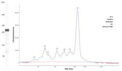
- Simple Western: Myoferlin Antibody [NBP1-84694] - Lane view shows a specific band for Myoferlin using 200 ug/ml of RT4 cell lysate and 4 ug/ml antibody dilution. Electropherogram image of corresponding Simple Western lane view at WES molecular weight of 222. Image from an internal validation.

- Submitted by
- Novus Biologicals (provider)
- Main image
- Experimental details
- Simple Western: Myoferlin Antibody [NBP1-84694] - Lane view shows a specific band for Myoferlin using 200 ug/mL of RT4 cell lysate and 4 ug/mL antibody dilution. Electropherogram image of corresponding Simple Western lane view at WES molecular weight of 222 kDa. Image from an internal validation.

Supportive validation
- Submitted by
- Novus Biologicals (provider)
- Main image

- Experimental details
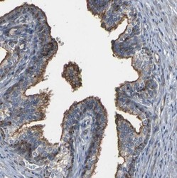
- Immunohistochemistry-Paraffin: Myoferlin Antibody [NBP1-84694] - Staining of human cervix, uterine shows high expression.

- Submitted by
- Novus Biologicals (provider)
- Main image

- Experimental details
- Immunohistochemistry-Paraffin: Myoferlin Antibody [NBP1-84694] - Staining of human testis shows low expression as expected.

- Submitted by
- Novus Biologicals (provider)
- Main image

- Experimental details
- Immunohistochemistry-Paraffin: Myoferlin Antibody [NBP1-84694] - Staining of human placenta shows moderate to strong membranous positivity in trophoblastic cells.

- Submitted by
- Novus Biologicals (provider)
- Main image
- Experimental details
- Immunohistochemistry-Paraffin: Myoferlin Antibody [NBP1-84694] - Staining of human prostate shows moderate to strong membranous positivity in glandular cells.

- Submitted by
- Novus Biologicals (provider)
- Main image

- Experimental details
- Immunohistochemistry-Paraffin: Myoferlin Antibody [NBP1-84694] - Staining of human skeletal muscle shows no positivity in myocytes as expected.

- Submitted by
- Novus Biologicals (provider)
- Main image

- Experimental details
- Immunohistochemistry-Paraffin: Myoferlin Antibody [NBP1-84694] - Staining of human urinary bladder shows strong membranous positivity in urothelial cells.

- Submitted by
- Novus Biologicals (provider)
- Main image

- Experimental details
- Immunohistochemistry-Paraffin: Myoferlin Antibody [NBP1-84694] - Staining in human urinary bladder and skeletal muscle tissues using NBP1-84694 antibody. Corresponding MYOF RNA-seq data are presented for the same tissues.
