Explore
 Validate
 Learn
Learn Western blot
 Immunocytochemistry
Antibody data
- Antibody Data
- Antigen structure
- References [1]
- Comments [0]
- Validations
- Immunocytochemistry [3]
- Immunohistochemistry [1]
- Other assay [1]
Submit
Validation data
Reference
Comment
Report error
- Product number
- PA5-72964 - Provider product page

- Provider
- Invitrogen Antibodies
- Product name
- CHD7 Polyclonal Antibody
- Antibody type
- Polyclonal
- Antigen
- Synthetic peptide
- Reactivity
- Human, Mouse
- Host
- Rabbit
- Isotype
- IgG
- Vial size
- 100 µL
- Concentration
- 1 mg/mL
- Storage
- Store at 4°C short term. For long term storage, store at -20°C, avoiding freeze/thaw cycles.
Submitted references The chromatin remodelling factor Chd7 protects auditory neurons and sensory hair cells from stress-induced degeneration.
Ahmed M, Moon R, Prajapati RS, James E, Basson MA, Streit A
Communications biology 2021 Nov 3;4(1):1260
Communications biology 2021 Nov 3;4(1):1260
No comments: Submit comment
Supportive validation
- Submitted by
- Invitrogen Antibodies (provider)
- Main image

- Experimental details
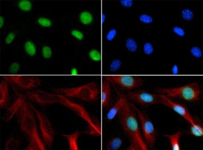
- Immunofluorescent analysis of NIH/3T3 cells using CHD7 polyclonal antibody (Product # PA5-72964) conjugated with FITC (green). Counterstain was performed with FITC (green), DAPI (blue) and Phalloidin (red).
- Submitted by
- Invitrogen Antibodies (provider)
- Main image

- Experimental details
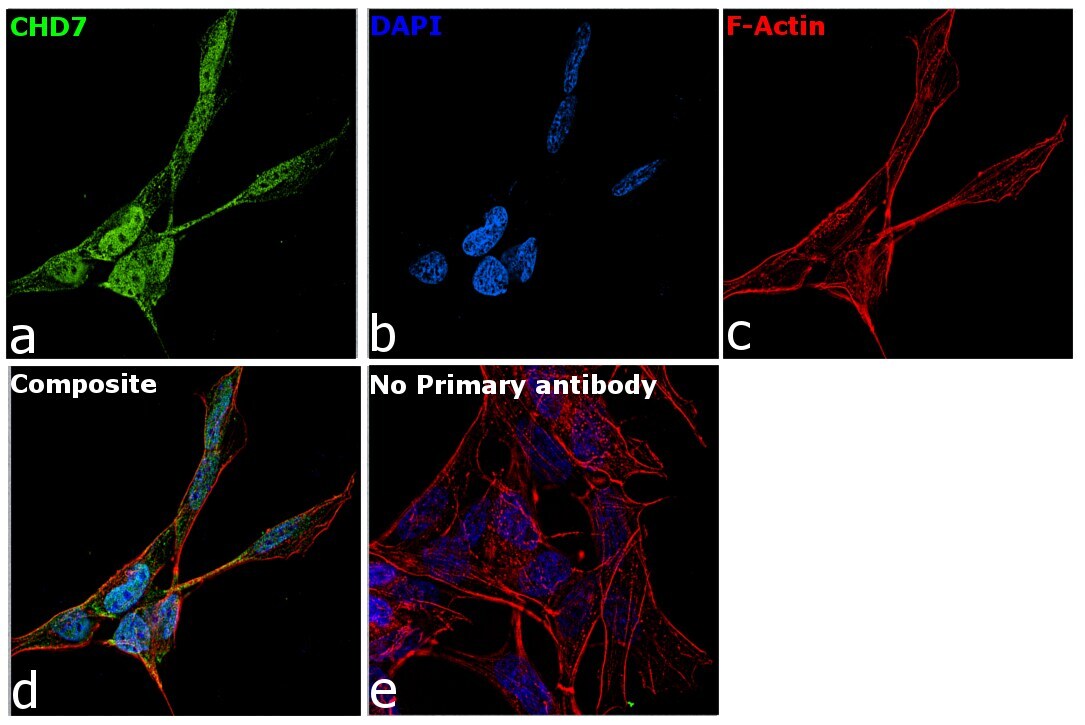
- Immunofluorescence analysis of CHD7 was performed using 70% confluent log phase SH-SY5Y cells. The cells were fixed with 4% paraformaldehyde for 10 minutes, permeabilized with 0.1% Triton™ X-100 for 15 minutes, and blocked with 2% BSA for 1 hour at room temperature. The cells were labeled with CHD7 Polyclonal Antibody (Product # PA5-72964) at 1:100 dilution in 0.1% BSA, incubated at 4 degree Celsius overnight and then labeled with Goat anti-Rabbit IgG (H+L) Superclonal™ Secondary Antibody, Alexa Fluor® 488 conjugate (Product # A27034) at a dilution of 1:2000 for 45 minutes at room temperature (Panel a: green). Nuclei (Panel b: blue) were stained with ProLong™ Diamond Antifade Mountant with DAPI (Product # P36962). F-actin (Panel c: red) was stained with Rhodamine Phalloidin (Product # R415, 1:300). Panel d represents the merged image showing nuclear localization. Panel e represents cells with no primary antibody to assess background. The images were captured at 60X magnification..
- Submitted by
- Invitrogen Antibodies (provider)
- Main image

- Experimental details
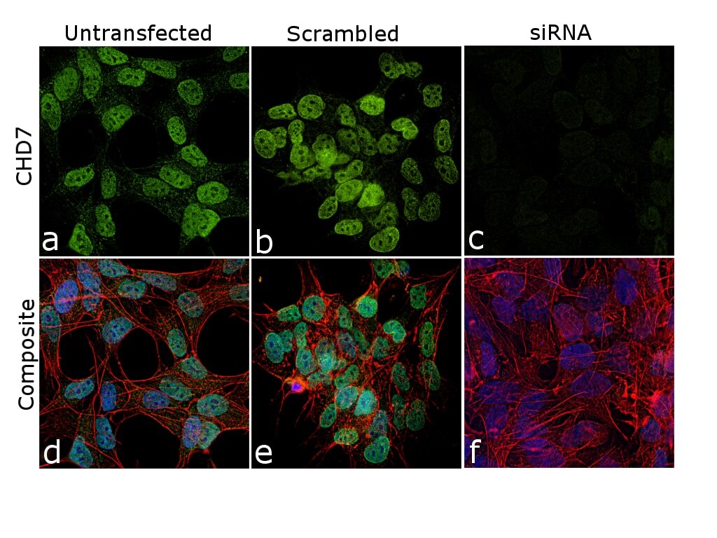
- KD of CHD7 was achieved by transfecting SH-SY5Y cells with CHD7 specific siRNA (Silencer® select Product # s31140, s529331). Immunofluorescence analysis was performed on untransfected SH-SY5Y cells (panel a,d), transfected with non-specific scrambled siRNA (panels b,e) and CHD7 specific siRNA (panel c,f). Cells were fixed, permeabilized, and labelled with CHD7 Polyclonal Antibody (Product # PA5-72964, 1:100 dilution), followed by Goat anti-Rabbit IgG (H+L) Superclonal™ Secondary Antibody, Alexa Fluor® 488 conjugate (Product # A27034, 1:2000). Nuclei (blue) were stained using ProLong™ Diamond Antifade Mountant with DAPI (Product # P36962), and Rhodamine Phalloidin (Product # R415, 1:300) was used for cytoskeletal F-actin (red) staining. Reduction of specific signal was observed upon siRNA mediated KD (panel c,f) confirming specificity of the antibody to CHD7. The images were captured at 60X magnification..
Supportive validation
- Submitted by
- Invitrogen Antibodies (provider)
- Main image
- Experimental details
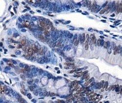
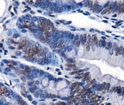
- Immunohistochemical analysis of CHD7 in mouse intestine. Samples were incubated with CHD7 polyclonal antibody (Product # PA5-72964) followed by using DAB with hematoxylin counterstain.
Supportive validation
- Submitted by
- Invitrogen Antibodies (provider)
- Main image

- Experimental details
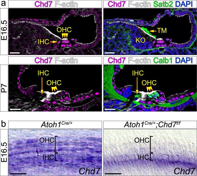
- Fig. 1 Chd7 expression in wildtype and Atoh1 Cre/+ ;Chd7 f/f mutant organ of Corti. a Immunohistochemistry in wildtype cochlea showing Chd7 expression in hair cells at E16.5 and P7. Expression is weaker at P7 compared to E16.5. Hair cells are stained with F-actin and Calbindin 1. Tectorial membrane is stained with Satb2. b In-situ hybridisation showing loss of Chd7 expression in Atoh1 Cre/+ ;Chd7 f/f mutant hair cells within the middle region of the cochlea at E16.5. Atoh1 Cre/+ or Chd7 floxed siblings were used as controls (indicated at the top of the panel). IHC inner hair cells; OHC outer hair cells; TM tectorial membrane. Scale bars 25 um.