 Explore
 Validate
 Learn
Learn Western blot
Antibody data
- Antibody Data
- Antigen structure
- References [1]
- Comments [0]
- Validations
- Western blot [3]
- Immunohistochemistry [1]
- Other assay [5]
Submit
Validation data
Reference
Comment
Report error
- Product number
- PA5-27994 - Provider product page

- Provider
- Invitrogen Antibodies
- Product name
- EDIL3 Polyclonal Antibody
- Antibody type
- Polyclonal
- Antigen
- Recombinant protein fragment
- Description
- Recommended positive controls: U87-MG conditioned medium, EDIL3-transfected 293T.
- Concentration
- 0.43 mg/mL
Submitted references EDIL3 promotes epithelial-mesenchymal transition and paclitaxel resistance through its interaction with integrin α(V)β(3) in cancer cells.
Gasca J, Flores ML, Jiménez-Guerrero R, Sáez ME, Barragán I, Ruíz-Borrego M, Tortolero M, Romero F, Sáez C, Japón MA
Cell death discovery 2020;6:86
Cell death discovery 2020;6:86
No comments: Submit comment
Supportive validation
- Submitted by
- Invitrogen Antibodies (provider)
- Main image

- Experimental details
- Western Blot analysis of EDIL3 was performed by separating 30 µg of non-transfected (–) and transfected (+) 293T whole cell extracts by 10% SDS-PAGE. Proteins were transferred to a membrane and probed with a EDIL3 Polyclonal Antibody (Product # PA5-27994) at a dilution of 1:10000. The HRP-conjugated anti-rabbit IgG antibody was used to detect the primary antibody.

- Submitted by
- Invitrogen Antibodies (provider)
- Main image
- Experimental details
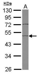
- Western Blot using EDIL3 Polyclonal Antibody (Product # PA5-27994). Sample (50 µg of whole cell lysate). Lane A: Mouse brain. 10% SDS PAGE. EDIL3 Polyclonal Antibody (Product # PA5-27994) diluted at 1:1,000. The HRP-conjugated anti-rabbit IgG antibody was used to detect the primary antibody.

- Submitted by
- Invitrogen Antibodies (provider)
- Main image

- Experimental details
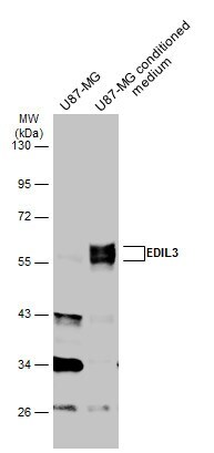
- Western blot analysis of EDIL3 was performed by separating 30 µg of u87-MG whole cell extract and conditioned medium by 10% SDS-PAGE. Proteins were transferred to a membrane and probed with a EDIL3 Polyclonal Antibody (Product # PA5-27994) at a dilution of 1:1000. The HRP-conjugated anti-rabbit IgG antibody was used to detect the primary antibody.
Supportive validation
- Submitted by
- Invitrogen Antibodies (provider)
- Main image

- Experimental details
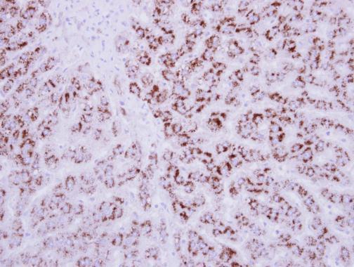
- Immunohistochemical analysis of paraffin-embedded human breast cancer, using EDIL3 (Product # PA5-27994) antibody at 1:250 dilution. Antigen Retrieval: EDTA based buffer, pH 8.0, 15 min.
Supportive validation
- Submitted by
- Invitrogen Antibodies (provider)
- Main image

- Experimental details
- Fig. 2 High EDIL3 protein expression is associated with a mesenchymal phenotype in paclitaxel-resistant cancer cells. Western blot analysis of basal expression of EDIL3, and different epithelial and mesenchymal markers are shown. beta-actin is shown as a loading control. Histograms show the densitometric analyses of indicated proteins. Data are presented as mean +- SEM ( n >= 3). a EDIL3 expression in MDA-MB-468, MDA-MB-468R, BT474, SKBR3, and MDA-MB-231 breast cancer cells. b E-cadherin, vimentin, and Slug expression in MDA-MB-468, MDA-MB-468R, and MDA-MB-231 breast cancer cells. c EDIL3, E-cadherin, vimentin, and Slug expression in LNCaP and PC3 prostate cancer cells. d , e Representative micrographs of EDIL3 immunohistochemistry in low- and high-grade breast tumors ( d ), and in low- and high-grade prostate tumors ( e ). Bars, 75 um. Histograms show percent of breast ( d ) or prostate ( e ) tumors with high EDIL3 expression. Association between EDIL3 and tumor grade was analyzed by Fisher's exact test. * P < 0.05 from Student's t test; NS not significant.

- Submitted by
- Invitrogen Antibodies (provider)
- Main image
- Experimental details
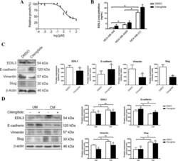
- Fig. 3 EDIL3 regulates epithelial-mesenchymal transition through an autocrine or paracrine mechanism in breast cancer cells. a IC50 curve for cilengitide in MDA-MB-231 breast cancer cell line. Data are presented as mean +- SEM. b Histogram shows the concentration of EDIL3 protein in the medium determined by ELISA in MDA-MB-468, MDA-MB-468R, and MDA-MB-231 breast cancer cells treated with DMSO or 1 uM cilengitide during 48 h. c Western blot analysis of EDIL3, E-cadherin, vimentin, and Slug in MDA-MB-231 cells treated with DMSO and 1 uM cilengitide during 48 h. beta-actin is shown as a loading control. Histograms show the densitometric analyses of indicated proteins. Data are presented as mean +- SEM ( n >= 3). d Western blot analysis of EDIL3, E-cadherin, vimentin, and Slug in MDA-MB-468 cells cultured in unconditioned medium (UM) or EDIL3-enriched conditioned medium collected from MDA-MB-231 cells (CM), and treated with DMSO and 1 uM cilengitide during 48 h. beta-actin is shown as a loading control. Histograms show the densitometric analyses of indicated proteins. Data are presented as mean +- SEM ( n >= 3). * P < 0.05 from Student's t test; NS not significant.

- Submitted by
- Invitrogen Antibodies (provider)
- Main image

- Experimental details
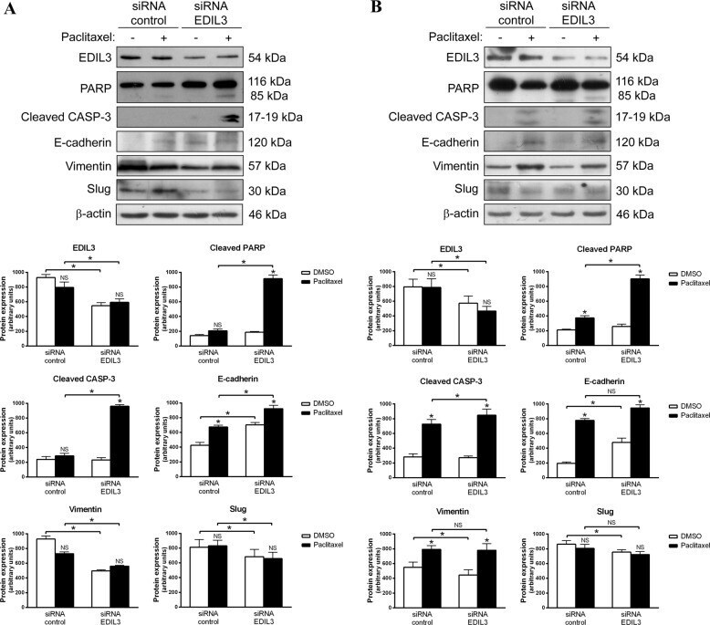
- Fig. 4 EDIL3 gene silencing increases paclitaxel-induced apoptosis and reverts epithelial-mesenchymal transition in paclitaxel-resistant cancer cells. a MDA-MB-231 and b PC3 cells were silenced for EDIL3, and treated with DMSO and 1 or 2.5 uM paclitaxel, respectively. Paclitaxel-induced apoptosis was assessed by western blot analysis of cleaved PARP and active caspase-3. Modulation of EDIL3, E-cadherin, vimentin, and Slug was studied by western blot analysis, using beta-actin as a loading control. Histograms show the densitometric analyses of indicated proteins. Data are presented as mean +- SEM ( n >= 3). * P < 0.05 from Student's t test; NS not significant.
- Submitted by
- Invitrogen Antibodies (provider)
- Main image

- Experimental details
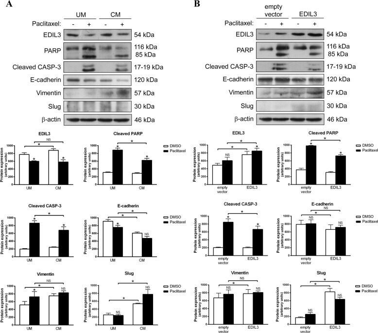
- Fig. 5 Extracellular EDIL3 and EDIL3 overexpression promotes epithelial-mesenchymal transition and paclitaxel resistance in cancer cells. a MDA-MB-468 breast cancer cells were cultured in unconditioned medium (UM) or EDIL3-enriched conditioned medium collected from MDA-MB-231 cells (CM), and treated with DMSO and 1 uM paclitaxel during 48 h. b LNCaP prostate cancer cells were transiently transfected with pCMV6-XL4-EDIL3 or with empty vector, and treated with DMSO or 2.5 uM paclitaxel during 48 h. Paclitaxel-induced cleavage of PARP and caspase-3, as well as paclitaxel-induced modulation of EDIL3, E-cadherin, vimentin, and Slug were analyzed by western blot using beta-actin as a loading control. Histograms represent the densitometric analyses of indicated proteins. Data are presented as mean +- SEM ( n >= 3). * P < 0.05 from Student's t test; NS, not significant.
- Submitted by
- Invitrogen Antibodies (provider)
- Main image

- Experimental details
- Fig. 6 Cilengitide sensitizes paclitaxel-resistant cancer cells to paclitaxel-induced apoptosis and reverts mesenchymal transition. a MDA-MB-231 and b PC3 cells were treated with DMSO, 1 uM cilengitide, 1 or 2.5 uM paclitaxel, and 1 uM cilengitide plus 1 or 2.5 uM paclitaxel, respectively. The induction of apoptosis was studied through the cleavage of PARP and caspase-3 by western blot, as well as the expression of indicated epithelial-mesenchymal transition markers. beta-actin was used as a loading control. Histograms show the densitometric analyses of indicated proteins. Data are presented as mean +- SEM ( n >= 3). * P < 0.05 from Student''s t test; NS not significant.
