Explore
 Validate
 Learn
Learn Western blot
Antibody data
- Antibody Data
- Antigen structure
- References [2]
- Comments [0]
- Validations
- Western blot [1]
- Immunohistochemistry [2]
- Other assay [1]
Submit
Validation data
Reference
Comment
Report error
- Product number
- MA5-27620 - Provider product page

- Provider
- Invitrogen Antibodies
- Product name
- TRPM7 Monoclonal Antibody (S74)
- Antibody type
- Monoclonal
- Antigen
- Other
- Reactivity
- Human, Mouse, Rat
- Host
- Mouse
- Isotype
- IgG
- Antibody clone number
- S74
- Vial size
- 100 µg
- Concentration
- 1 mg/mL
- Storage
- -20°C
Submitted references The molecular appearance of native TRPM7 channel complexes identified by high-resolution proteomics.
Epidermal growth factor signaling through transient receptor potential melastatin 7 cation channel regulates vascular smooth muscle cell function.
Kollewe A, Chubanov V, Tseung FT, Correia L, Schmidt E, Rössig A, Zierler S, Haupt A, Müller CS, Bildl W, Schulte U, Nicke A, Fakler B, Gudermann T
eLife 2021 Nov 12;10
eLife 2021 Nov 12;10
Epidermal growth factor signaling through transient receptor potential melastatin 7 cation channel regulates vascular smooth muscle cell function.
Zou ZG, Rios FJ, Neves KB, Alves-Lopes R, Ling J, Baillie GS, Gao X, Fuller W, Camargo LL, Gudermann T, Chubanov V, Montezano AC, Touyz RM
Clinical science (London, England : 1979) 2020 Aug 14;134(15):2019-2035
Clinical science (London, England : 1979) 2020 Aug 14;134(15):2019-2035
No comments: Submit comment
Supportive validation
- Submitted by
- Invitrogen Antibodies (provider)
- Main image
- Experimental details
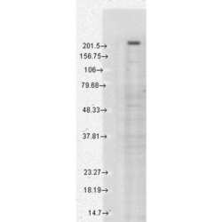
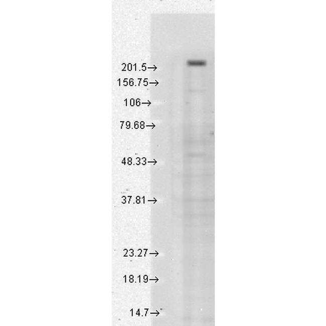
- Western blot analysis of TRPM7 in human cell lysates with 15 µg of sample. The sample was blocked with 1.5% BSA, incubated with TRPM7 monoclonal antibody (Product # MA5-27620) using a dilution of 1:1000 (2 hours at 4°C), followed by Sheep Anti-Mouse HRP (1 hour at RT).
Supportive validation
- Submitted by
- Invitrogen Antibodies (provider)
- Main image
- Experimental details
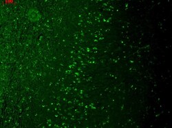
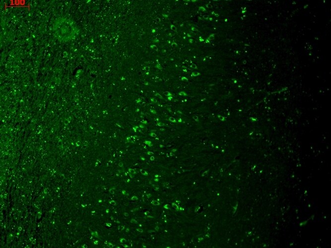
- Immunohistochemistry analysis of TRPM7 in paraffin-embedded human hippocampus. Sample was fixed with Bouin's Fixative Solution, incubated with TRPM7 monoclonal antibody (Product # MA5-27620) using a dilution of 1:1000 (1 hour at RT), and followed by FITC Goat Anti-Mouse (green) (1 hour at RT) at a dilution of 1:50.
- Submitted by
- Invitrogen Antibodies (provider)
- Main image
- Experimental details
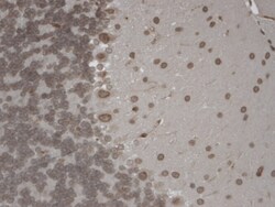
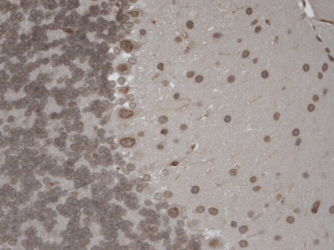
- Immunohistochemistry analysis of TRPM7 in paraffin-embedded mouse hippocampus. Samples were incubated with TRPM7 monoclonal antibody (Product # MA5-27620) using a dilution of 1:1000 (1 hour at RT) and followed by Biotinylated Goat Anti-Mouse, Streptavidin Peroxidase, DAB Chromogen (brown) and Mayer Hematoxylin (purple/blue) at a dilution of 250-500 µl.
Supportive validation
- Submitted by
- Invitrogen Antibodies (provider)
- Main image
- Experimental details
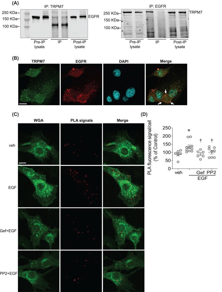
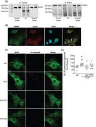
- Figure 2 EGF enhances direct interaction between EGFR and TRPM7 in a c-Src-dependent manner ( A ) EGFR was detected when TRPM7 was immunoprecipitated in rVSMCs using anti-TRPM7 antibody (left panel) and TRPM7 was detected when EGFR was immunoprecipitated using anti-EGFR antibody (right panel) ( n =3-4). ( B ) Representative confocal microscopy images of rVSMCs co-immunostained for TRPM7 (Alexa 488, green) and EGFR (Alexa 555, red). Nuclei were stained with DAPI ( n =3). ( C , D ) PLA was used to visualize and quantify TRPM7-EGFR interaction in rVSMCs stimulated with vehicle (veh), and EGF (50 ng/ml) in the presence and absence of gefitinib (1 uM) and PP2 (10 uM). WGA (Alexa 488, green) was used to stain cell membranes. Red fluorescence is identified as PLA positive signals. Orange fluorescence in the merged figure identifies co-localization. PLA signals were quantified using the Analyse Particles plugin of ImageJ software. For each condition, quantifications were performed from at least 100 cells and expressed as the mean number of signals per cell (percentage of control). Scale bar =10 um. Arrows indicate co-localization of EGFR and TRPM7. * P