Explore
 Validate
 Learn
Learn Western blot
 Immunocytochemistry
Antibody data
- Antibody Data
- Antigen structure
- References [3]
- Comments [0]
- Validations
- Immunocytochemistry [1]
- Immunohistochemistry [6]
Submit
Validation data
Reference
Comment
Report error
- Product number
- HPA012897 - Provider product page

- Provider
- Atlas Antibodies
- Proper citation
- Atlas Antibodies Cat#HPA012897, RRID:AB_1855949
- Product name
- Anti-PTRH2
- Antibody type
- Polyclonal
- Reactivity
- Human
- Host
- Rabbit
- Conjugate
- Unconjugated
- Antigen sequence
KSKTSKTHTDTESEASILGDSGEYKMILVVRNDLK
MGKGKVAAQCSHAAVSAYKQIQRRNPEMLKQWEYC
GQPKVVVKAPDEETLIALLAHAKMLGLTVSLIQDA
GRTQIAPGSQTVLGIGPGPADLIDKVTGHLKL- Isotype
- IgG
- Vial size
- 100 µl
- Storage
- Store at +4°C for short term storage. Long time storage is recommended at -20°C.
Submitted references The anoikis effector Bit1 displays tumor suppressive function in lung cancer cells.
TLE1 is an anoikis regulator and is downregulated by Bit1 in breast cancer cells.
Metastasis of tumor cells is enhanced by downregulation of Bit1.
Yao X, Jennings S, Ireland SK, Pham T, Temple B, Davis M, Chen R, Davenport I, Biliran H
PloS one 2014;9(7):e101564
PloS one 2014;9(7):e101564
TLE1 is an anoikis regulator and is downregulated by Bit1 in breast cancer cells.
Brunquell C, Biliran H, Jennings S, Ireland SK, Chen R, Ruoslahti E
Molecular cancer research : MCR 2012 Nov;10(11):1482-95
Molecular cancer research : MCR 2012 Nov;10(11):1482-95
Metastasis of tumor cells is enhanced by downregulation of Bit1.
Karmali PP, Brunquell C, Tram H, Ireland SK, Ruoslahti E, Biliran H
PloS one 2011;6(8):e23840
PloS one 2011;6(8):e23840
No comments: Submit comment
Supportive validation
- Submitted by
- Atlas Antibodies (provider)
- Main image
- Experimental details
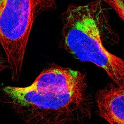
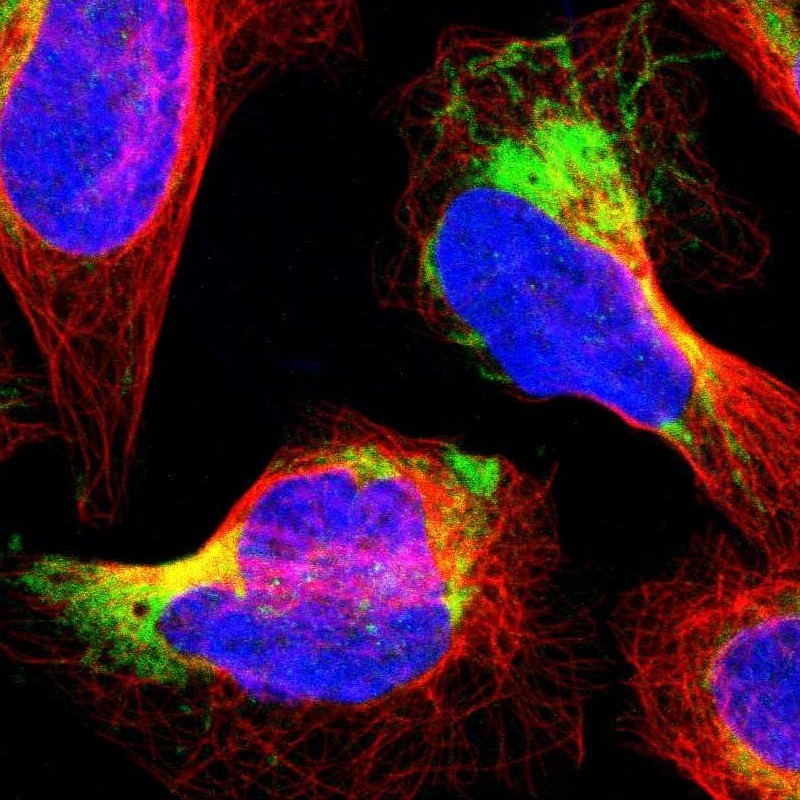
- Immunofluorescent staining of human cell line U-2 OS shows localization to mitochondria.
- Sample type
- HUMAN
Supportive validation
- Submitted by
- Atlas Antibodies (provider)
- Main image
- Experimental details
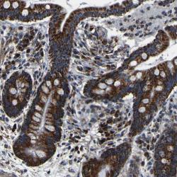
- Immunohistochemical staining of human colon shows strong cytoplasmic positivity, with a granular pattern, in glandular cells.
- Sample type
- HUMAN
- Submitted by
- Atlas Antibodies (provider)
- Main image
- Experimental details
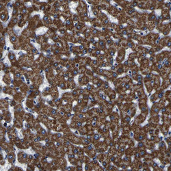
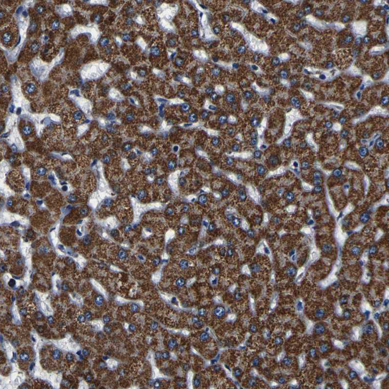
- Immunohistochemical staining of human liver shows strong granular cytoplasmic positivity in hepatocytes.
- Sample type
- HUMAN
- Submitted by
- Atlas Antibodies (provider)
- Main image
- Experimental details
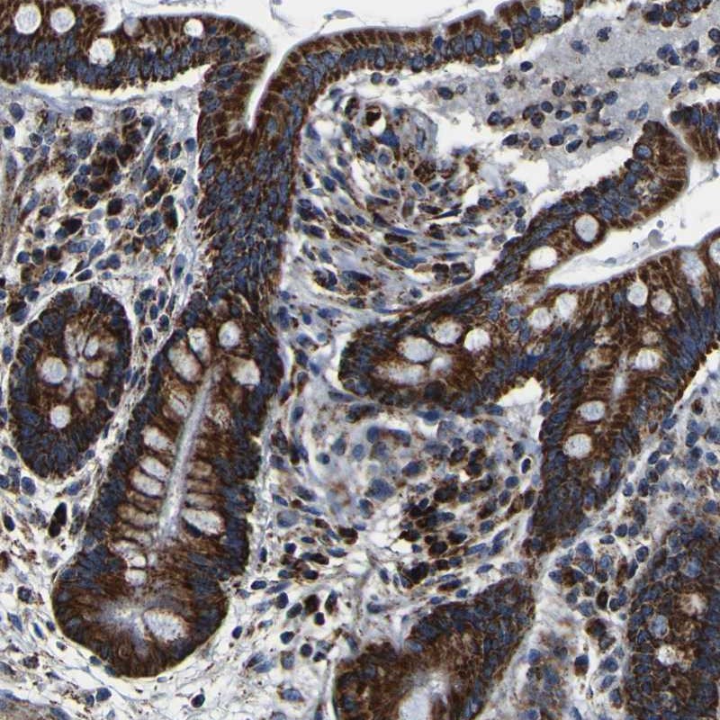
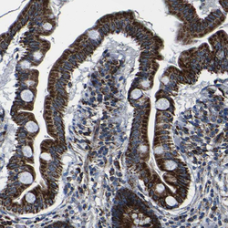
- Immunohistochemical staining of human small intestine shows strong granular cytoplasmic positivity in glandular cells.
- Sample type
- HUMAN

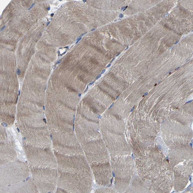
- Submitted by
- Atlas Antibodies (provider)
- Main image

- Experimental details
- Immunohistochemical staining of human skeletal muscle shows moderate cytoplasmic positivity in myocytes.
- Sample type
- HUMAN
- Submitted by
- Atlas Antibodies (provider)
- Main image
- Experimental details
- Immunohistochemical staining of human testis shows strong granular cytoplasmic positivity in cells in seminiferous ducts.
- Sample type
- HUMAN

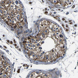
- Submitted by
- Atlas Antibodies (provider)
- Main image

- Experimental details
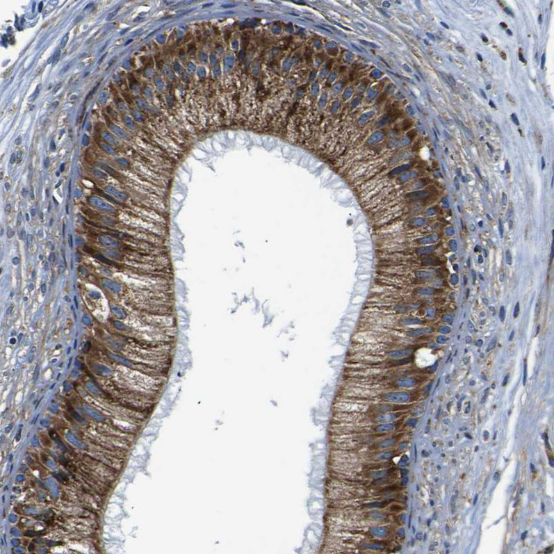
- Immunohistochemical staining of human epididymis shows strong cytoplasmic positivity in glandular cells.
- Sample type
- HUMAN