Explore
 Validate
 Learn
Learn Western blot
 Immunohistochemistry
Antibody data
- Antibody Data
- Antigen structure
- References [1]
- Comments [0]
- Validations
- Immunohistochemistry [1]
Submit
Validation data
Reference
Comment
Report error
- Product number
- AF3495 - Provider product page

- Provider
- R&D Systems
- Product name
- Human LRIG3 Antibody
- Antibody type
- Polyclonal
- Description
- Antigen Affinity-purified. Detects human LRIG3 in direct ELISAs and Western blots. In direct ELISAs, less than 1% cross-reactivity with recombinant mouse LRIG1 is observed.
- Reactivity
- Human
- Host
- Goat
- Conjugate
- Unconjugated
- Antigen sequence
Q6UXM1- Isotype
- IgG
- Vial size
- 100 ug
- Concentration
- LYOPH
- Storage
- Use a manual defrost freezer and avoid repeated freeze-thaw cycles. 12 months from date of receipt, -20 to -70 °C as supplied. 1 month, 2 to 8 °C under sterile conditions after reconstitution. 6 months, -20 to -70 °C under sterile conditions after reconstitution.
Submitted references Leucine-rich repeat and immunoglobulin domain-containing protein-1 (Lrig1) negative regulatory action toward ErbB receptor tyrosine kinases is opposed by leucine-rich repeat and immunoglobulin domain-containing protein 3 (Lrig3).
Rafidi H, Mercado F 3rd, Astudillo M, Fry WH, Saldana M, Carraway KL 3rd, Sweeney C
The Journal of biological chemistry 2013 Jul 26;288(30):21593-605
The Journal of biological chemistry 2013 Jul 26;288(30):21593-605
No comments: Submit comment
Supportive validation
- Submitted by
- R&D Systems (provider)
- Main image
- Experimental details
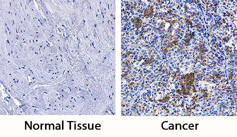
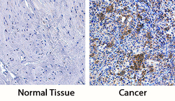
- LRIG3 in Human Cervical Cancer Tissue. LRIG3 was detected in immersion fixed paraffin-embedded sections of human cervical cancer tissue using Goat Anti-Human LRIG3 Antigen Affinity-purified Polyclonal Antibody (Catalog # AF3495) at 3 µg/mL overnight at 4 °C. Tissue was stained using the Anti-Goat HRP-DAB Cell & Tissue Staining Kit (brown; Catalog # CTS008) and counterstained with hematoxylin (blue). Specific staining was localized to cytoplasm of cancer cells. View our protocol for Chromogenic IHC Staining of Paraffin-embedded Tissue Sections.