 Explore
 Validate
 Learn
Learn Western blot
 Immunocytochemistry
Antibody data
- Antibody Data
- Antigen structure
- References [2]
- Comments [0]
- Validations
- Immunocytochemistry [1]
- Immunohistochemistry [1]
Submit
Validation data
Reference
Comment
Report error
- Product number
- GTX22873 - Provider product page

- Provider
- GeneTex
- Proper citation
- GeneTex Cat#GTX22873, RRID:AB_384892
- Product name
- Sodium/Potassium ATPase beta 1 antibody [M17-P5-F11]
- Antibody type
- Monoclonal
- Reactivity
- Human, Mouse, Canine, Drosophila, Porcine, Sheep
- Host
- Mouse
Submitted references Dystrophin Threshold Level Necessary for Normalization of Neuronal Nitric Oxide Synthase, Inducible Nitric Oxide Synthase, and Ryanodine Receptor-Calcium Release Channel Type 1 Nitrosylation in Golden Retriever Muscular Dystrophy Dystrophinopathy.
Sodium channels contribute to degeneration of dorsal root ganglion neurites induced by mitochondrial dysfunction in an in vitro model of axonal injury.
Gentil C, Le Guiner C, Falcone S, Hogrel JY, Peccate C, Lorain S, Benkhelifa-Ziyyat S, Guigand L, Montus M, Servais L, Voit T, Piétri-Rouxel F
Human gene therapy 2016 Sep;27(9):712-26
Human gene therapy 2016 Sep;27(9):712-26
Sodium channels contribute to degeneration of dorsal root ganglion neurites induced by mitochondrial dysfunction in an in vitro model of axonal injury.
Persson AK, Kim I, Zhao P, Estacion M, Black JA, Waxman SG
The Journal of neuroscience : the official journal of the Society for Neuroscience 2013 Dec 4;33(49):19250-61
The Journal of neuroscience : the official journal of the Society for Neuroscience 2013 Dec 4;33(49):19250-61
No comments: Submit comment
Supportive validation
- Submitted by
- GeneTex (provider)
- Main image
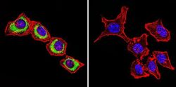
- Experimental details
- Immunofluorescent analysis of Sodium/Potassium ATPase beta in HeLa cells. Sodium/Potassium ATPase beta staining (green), F-Actin staining with Phalloidin (red) and nuclei with DAPI (blue) is shown. Cells were grown on slides and fixed with formaldehyde prior to staining. Cells were probed without (control) or with Sodium/Potassium ATPase beta antibody [M17-P5-F11] at a dilution of 1:200 over night at 4?C, washed with PBS and incubated with a proper secondary antibody. Images were taken at 60X magnification.

Supportive validation
- Submitted by
- GeneTex (provider)
- Main image

- Experimental details
- Immunohistochemistry was performed on cancer biopsies of deparaffinized human colon carcinoma tissues. To expose target proteins, heat induced antigen retrieval was performed using 10mM sodium citrate (pH6.0) buffer, microwaved for 8-15 minutes. Following antigen retrieval tissues were blocked in 3% BSA-PBS for 30 minutes at room temperature. Tissues were then probed at a dilution of 1:200 with or without Sodium/Potassium ATPase beta antibody [M17-P5-F11] overnight at 4¢XC in a humidified chamber. Tissues were washed extensively with PBST and endogenous peroxidase activity was quenched with a peroxidase suppressor. Detection was performed using a biotin-conjugated secondary antibody and SA-HRP, followed by colorimetric detection using DAB. Tissues were counterstained with hematoxylin and prepped for mounting.
