 Explore
 Validate
 Learn
Learn Western blot
Antibody data
- Antibody Data
- Antigen structure
- References [0]
- Comments [0]
- Validations
- Western blot [4]
- Immunohistochemistry [1]
- Other assay [1]
Submit
Validation data
Reference
Comment
Report error
- Product number
- PA5-47959 - Provider product page

- Provider
- Invitrogen Antibodies
- Product name
- Contactin 1 Polyclonal Antibody
- Antibody type
- Polyclonal
- Antigen
- Recombinant full-length protein
- Description
- Reconstitute at 0.2 mg/mL in sterile PBS.
- Concentration
- 0.2 mg/mL
No comments: Submit comment
Supportive validation
- Submitted by
- Invitrogen Antibodies (provider)
- Main image

- Experimental details
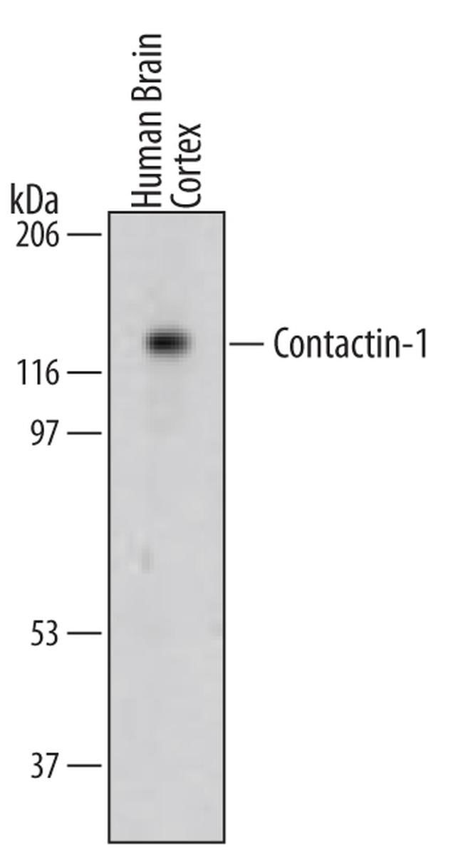
- Western blot analysis from lysates of human brain cortex tissue. PVDF membrane was probed with 1 µg/mL of Goat Anti-human Contactin-1 Antigen Affinity-purified Polyclonal Antibody (Product # PA5-47959) followed by HRP-conjugated Anti-Goat IgG Secondary Antibody. A specific band was detected for Contactin-1 at approximately 118 kDa (as indicated). This experiment was conducted under reducing conditions.
- Submitted by
- Invitrogen Antibodies (provider)
- Main image

- Experimental details
- Western blot analysis of Contactin 1 in human brain (cortex) tissue, human brain (cerebellum) tissue, mouse brain (cerebellum) tissue, mouse brain (total) tissue, and rat brain (total) tissue. Samples were incubated in Contactin 1 polyclonal antibody (Product # PA5-47959) using a dilution of 0.5 µg/mL followed by a HRP-conjugated Anti-Goat IgG secondary antibody. A specific band was detected for Contactin‚1 at approximately 135 kDa (as indicated). This experiment was conducted under reducing conditions.

- Submitted by
- Invitrogen Antibodies (provider)
- Main image

- Experimental details
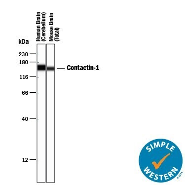
- Western blot analysis of Contactin 1 in 0.2 mg/mL lysates of human brain tissue and mouse brain tissue. Samples were incubated in Contactin 1 polyclonal antibody (Product # PA5-47959) using a dilution of 5 µg/mL followed by HRP-conjugated Anti-Goat IgG at a dilution of 1:50. A specific band was detected for Contactin‚1 at approximately 158 kDa (as indicated). This experiment was conducted under reducing conditions and using the 12-230 kDa separation system.
Supportive validation
- Submitted by
- Invitrogen Antibodies (provider)
- Main image

- Experimental details
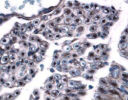
- Immunohistochemical analysis of Contactin 1 in immersion fixed paraffin-embedded sections of human dorsal root ganglia. Samples were incubated in Contactin 1 polyclonal antibody (Product # PA5-47959) using a dilution of 15 µg/mL overnight at 4 °C. Tissue was stained with the Anti-Goat HRP-DAB Cell & Tissue Staining Kit (brown) and counterstained with hematoxylin (blue).
Supportive validation
- Submitted by
- Invitrogen Antibodies (provider)
- Main image

- Experimental details
- Neutralization of Contactin 1 in C6 rat glioma cell line. Samples were incubated in Contactin 1 polyclonal antibody (Product # PA5-47959). Recombinant Human Contactin‚1 Fc Chimera, immobilized onto a microplate, supports the adhesion of the C6 rat glioma cell line in a dose-dependent manner (orange line). Adhesion elicited by Recombinant Human Contactin‚1 Fc Chimera (2 µg/mL) is neutralized (green line) by increasing concentrations of Goat Anti-Human/Mouse/RatContactin‚1 Antigen Affinity-purified Polyclonal Antibody. The ND50 is typically 1-5 µg/mL.
