Explore
 Validate
 Learn
Learn Western blot
Antibody data
- Antibody Data
- Antigen structure
- References [0]
- Comments [0]
- Validations
- Western blot [4]
- Immunocytochemistry [1]
- Immunohistochemistry [2]
Submit
Validation data
Reference
Comment
Report error
- Product number
- GTX129202 - Provider product page

- Provider
- GeneTex
- Proper citation
- GeneTex Cat#GTX129202, RRID:AB_2797598
- Product name
- ER81 antibody
- Antibody type
- Polyclonal
- Reactivity
- Human, Mouse, Rat
- Host
- Rabbit
No comments: Submit comment
Supportive validation
- Submitted by
- GeneTex (provider)
- Main image

- Experimental details
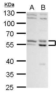
- ER81 antibody detects ER81 protein by western blot analysis.A GL261 whole cell lysate/extractB C8D30 whole cell lysate/extract10 % SDS-PAGEER81 antibody (GTX129202) dilution: 1:1000
- Validation comment
- WB
- Submitted by
- GeneTex (provider)
- Main image

- Experimental details
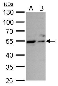
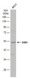
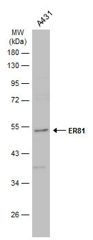
- ER81 antibody detects ER81 protein by western blot analysis.A. 30 ?g A431 whole cell lysate/extractB. 30 ?g HeLa whole cell lysate/extract10% SDS-PAGEER81 antibody (GTX129202) dilution: 1:1000 The HRP-conjugated anti-rabbit IgG antibody (GTX213110-01) was used to detect the primary antibody.
- Submitted by
- GeneTex (provider)
- Main image

- Experimental details
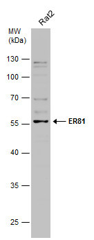
- Whole cell extract (30 ?g) was separated by 10% SDS-PAGE, and the membrane was blotted with ER81 antibody (GTX129202) diluted at 1:500. The HRP-conjugated anti-rabbit IgG antibody (GTX213110-01) was used to detect the primary antibody.
- Submitted by
- GeneTex (provider)
- Main image
- Experimental details
- Whole cell extract (30 ?g) was separated by 10% SDS-PAGE, and the membrane was blotted with ER81 antibody (GTX129202) diluted at 1:1000. The HRP-conjugated anti-rabbit IgG antibody (GTX213110-01) was used to detect the primary antibody.
Supportive validation
- Submitted by
- GeneTex (provider)
- Main image
- Experimental details
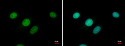
- ER81 antibody detects ER81 protein at nucleus by immunofluorescent analysis.Sample: C8D30 cells were fixed in 4% paraformaldehyde at RT for 15 min.Green: ER81 protein stained by ER81 antibody (GTX129202) diluted at 1:500.Blue: Hoechst 33342 staining.

Supportive validation
- Submitted by
- GeneTex (provider)
- Main image
- Experimental details
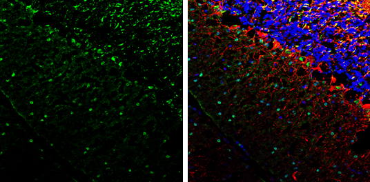
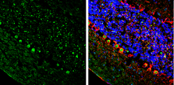
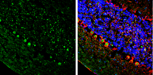
- ER81 antibody detects ER81 protein by immunohistochemical analysis.Sample: Frozen-sectioned mouse cerebellum.Green: ER81 stained by ER81 antibody (GTX129202) diluted at 1:250.Red: NF-H, stained by NF-H antibody [GT114] (GTX634289) diluted at 1:500.Blue: Fluoroshield with DAPI (GTX30920).
- Submitted by
- GeneTex (provider)
- Main image
- Experimental details
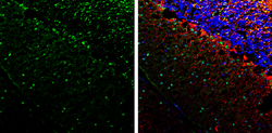
- ER81 antibody detects ER81 protein by immunohistochemical analysis.Sample: Frozen-sectioned mouse cerebellum.Green: ER81 stained by ER81 antibody (GTX129202) diluted at 1:250.Red: NF-H, stained by NF-H antibody [GT114] (GTX634289) diluted at 1:500.Blue: Fluoroshield with DAPI (GTX30920).