Antibody data
- Antibody Data
- Antigen structure
- References [4]
- Comments [0]
- Validations
- Western blot [3]
Submit
Validation data
Reference
Comment
Report error
- Product number
- NB100-2268 - Provider product page

- Provider
- Novus Biologicals
- Proper citation
- Novus Cat#NB100-2268, RRID:AB_2182048
- Product name
- Rabbit Polyclonal RPL7 Antibody
- Antibody type
- Polyclonal
- Description
- Immunogen affinity purified.
- Reactivity
- Human, Mouse
- Host
- Rabbit
- Isotype
- IgG
- Vial size
- 100 ul
- Concentration
- 1.0 mg/ml
- Storage
- Store at 4C. Do not freeze.
Submitted references The ribosomal protein Rpl22 controls ribosome composition by directly repressing expression of its own paralog, Rpl22l1.
Characterization of staufen1 ribonucleoproteins by mass spectrometry and biochemical analyses reveal the presence of diverse host proteins associated with human immunodeficiency virus type 1.
Cell-type-specific isolation of ribosome-associated mRNA from complex tissues.
Nucleophosmin serves as a rate-limiting nuclear export chaperone for the Mammalian ribosome.
O'Leary MN, Schreiber KH, Zhang Y, Duc AC, Rao S, Hale JS, Academia EC, Shah SR, Morton JF, Holstein CA, Martin DB, Kaeberlein M, Ladiges WC, Fink PJ, Mackay VL, Wiest DL, Kennedy BK
PLoS genetics 2013;9(8):e1003708
PLoS genetics 2013;9(8):e1003708
Characterization of staufen1 ribonucleoproteins by mass spectrometry and biochemical analyses reveal the presence of diverse host proteins associated with human immunodeficiency virus type 1.
Milev MP, Ravichandran M, Khan MF, Schriemer DC, Mouland AJ
Frontiers in microbiology 2012;3:367
Frontiers in microbiology 2012;3:367
Cell-type-specific isolation of ribosome-associated mRNA from complex tissues.
Sanz E, Yang L, Su T, Morris DR, McKnight GS, Amieux PS
Proceedings of the National Academy of Sciences of the United States of America 2009 Aug 18;106(33):13939-44
Proceedings of the National Academy of Sciences of the United States of America 2009 Aug 18;106(33):13939-44
Nucleophosmin serves as a rate-limiting nuclear export chaperone for the Mammalian ribosome.
Maggi LB Jr, Kuchenruether M, Dadey DY, Schwope RM, Grisendi S, Townsend RR, Pandolfi PP, Weber JD
Molecular and cellular biology 2008 Dec;28(23):7050-65
Molecular and cellular biology 2008 Dec;28(23):7050-65
No comments: Submit comment
Supportive validation
- Submitted by
- Novus Biologicals (provider)
- Main image

- Experimental details
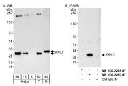
- Western Blot: RPL7 Antibody [NB100-2268] - Detection of Human and Mouse RPL7 on HeLa whole cell lysate using NB100-2268. RPL7 was immunoprecipitated by rabbit anti-RPL7 antibody NB100-2269.
- Submitted by
- Novus Biologicals (provider)
- Main image

- Experimental details
- Western Blot: RPL7 Antibody [NB100-2268] - Staufen1 co-fractionates with Gag and vRNA in gradient density fractionation analyses. HeLa cells were either mock transfected with empty vector, pcDNA3, or with a plasmid that expresses Staufen1-HA. The transfected cells were collected 24?h later, lysed and were either mock-treated or treated with RNAse A. The lysates were then fractionated on 5-50% sucrose gradients and 20 fractions were collected for further analysis by western blotting for viral and host proteins, as indicated. HIV-1 viral genomic RNA (vRNA, 9?kb) was assessed in each fraction by slot blot analysis. TL represents the total lysates. Image collected and cropped by CiteAb from the following publication (http://www.frontiersin.org/Journal/10.3389/fmicb.2012.00367/full), licensed under a CC-BY licence.
- Submitted by
- Novus Biologicals (provider)
- Main image

- Experimental details
- Western Blot: RPL7 Antibody [NB100-2268] - Staufen1 co-fractionates with Gag and vRNA in gradient density fractionation analyses. HeLa cells were co-transfected with pNL 4-3 and Staufen1-HA. The presence of Staufen1-HA, precursor Gag and p24 were assessed in each fraction by western blotting analysis. HIV-1 viral genomic RNA (9?kb, vRNA) was assessed in each fraction by slot blot analysis. Staufen1, Gag and vRNA were quantitated in each fraction by densitometry and relative levels are depicted for each fraction (Blue: Staufen1-HA, Red: Gag, Green-vRNA). Image collected and cropped by CiteAb from the following publication (http://www.frontiersin.org/Journal/10.3389/fmicb.2012.00367/full), licensed under a CC-BY licence.
 Explore
 Validate
 Learn
Learn Western blot
 Immunoprecipitation