Explore
 Validate
 Learn
Learn Western blot
Antibody data
- Antibody Data
- Antigen structure
- References [1]
- Comments [0]
- Validations
- Western blot [4]
- Immunoprecipitation [1]
Submit
Validation data
Reference
Comment
Report error
- Product number
- NBP1-52650 - Provider product page

- Provider
- Novus Biologicals
- Proper citation
- Novus Cat#NBP1-52650, RRID:AB_11053025
- Product name
- Rabbit Polyclonal E2F8 Antibody
- Antibody type
- Polyclonal
- Description
- Immunogen affinity purified.
- Reactivity
- Human, Mouse
- Host
- Rabbit
- Isotype
- IgG
- Vial size
- 100 ul
- Concentration
- 1.0 mg/ml
- Storage
- Store at 4C. Do not freeze.
Submitted references Histone deacetylase 11 inhibition promotes breast cancer metastasis from lymph nodes.
Leslie PL, Chao YL, Tsai YH, Ghosh SK, Porrello A, Van Swearingen AED, Harrison EB, Cooley BC, Parker JS, Carey LA, Pecot CV
Nature communications 2019 Sep 13;10(1):4192
Nature communications 2019 Sep 13;10(1):4192
No comments: Submit comment
Supportive validation
- Submitted by
- Novus Biologicals (provider)
- Main image

- Experimental details
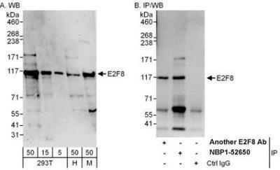
- Western Blot: E2F8 Antibody [NBP1-52650] - Whole cell lysate from HeLa (H; 50 mcg for WB; 1 mg for IP, 20% of IP loaded), 293T (5, 15, and 50 mcg for WB) and mouse NIH3T3 (M; 50 mcg) cells. Affinity purified rabbit anti-E2F8 antibody used for WB at 0.1 mcg/ml (A) and 1 mcg/ml (B) and used for IP at 6 mcg/mg lysate. E2F8 was also immunoprecipitated by another rabbit anti-E2F8 antibody, which recognizes an upstream epitope.
- Submitted by
- Novus Biologicals (provider)
- Main image

- Experimental details
- Western Blot: E2F8 Antibody [NBP1-52650] - Whole cell lysate (15 ug) from HeLa, 293T, and Jurkat cells prepared using NETN lysis buffer. Antibody: Affinity purified rabbit anti-E2F8 antibody used for WB at 0.1 ug/ml. Detection: Chemiluminescence with an exposure time of 30 seconds.

- Submitted by
- Novus Biologicals (provider)
- Main image
- Experimental details
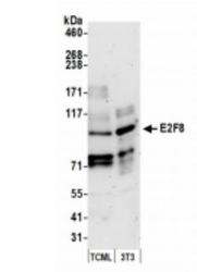
- Western Blot: E2F8 Antibody [NBP1-52650] - Whole cell lysate (15 ug) from TCMK-1 and NIH3T3 cells prepared using NETN lysis buffer. Antibody: Affinity purified rabbit anti-E2F8 antibody used for WB at 0.1 ug/ml. Detection: Chemiluminescence with an exposure time of 3 minutes.

- Submitted by
- Novus Biologicals (provider)
- Main image

- Experimental details
- Western Blot: E2F8 Antibody [NBP1-52650] - Detection of human E2F8 by western blot. Samples: Whole cell lysate (10 ug) from HEK293T and Jurkat cells prepared using NETN lysis buffer. Antibody: Affinity purified rabbit anti-E2F8 antibody NBP1-52650 used for WB at 0.1 ug/ml. Detection: Chemiluminescence with an exposure time of 3 minutes.

Supportive validation
- Submitted by
- Novus Biologicals (provider)
- Main image

- Experimental details
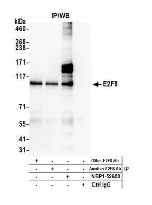
- Immunoprecipitation: E2F8 Antibody [NBP1-52650] - Detection of human E2F8 by western blot of immunoprecipitates. Samples: Whole cell lysate (1.0 mg per IP reaction; 20% of IP loaded) from HeLa cells prepared using NETN lysis buffer. Antibodies: Affinity purified rabbit anti-E2F8 antibody NBP1-52650 used for IP at 3 ug per reaction. E2F8 was also immunoprecipitated by two other rabbit anti-E2F8 antibodies. For blotting immunoprecipitated E2F8, NBP1-52650 was used at 1 ug/ml. Detection: Chemiluminescence with an exposure time of 3 minutes.