Explore
 Validate
 Learn
Learn Western blot
Antibody data
- Antibody Data
- Antigen structure
- References [3]
- Comments [0]
- Validations
- Western blot [2]
- Immunohistochemistry [1]
Submit
Validation data
Reference
Comment
Report error
- Product number
- AF5857 - Provider product page

- Provider
- R&D Systems
- Product name
- Human/Mouse NKX6.1 Antibody
- Antibody type
- Polyclonal
- Description
- Antigen Affinity-purified. Detects human and mouse NKX6.1 in direct ELISAs and Western blots. In direct ELISAs, less than 1% cross-reactivity with recombinant human NKX3.1 is observed.
- Reactivity
- Human, Mouse
- Host
- Goat
- Conjugate
- Unconjugated
- Antigen sequence
NP_006159- Isotype
- IgG
- Vial size
- 100 ug
- Concentration
- LYOPH
- Storage
- Use a manual defrost freezer and avoid repeated freeze-thaw cycles. 12 months from date of receipt, -20 to -70 °C as supplied. 1 month, 2 to 8 °C under sterile conditions after reconstitution. 6 months, -20 to -70 °C under sterile conditions after reconstitution.
Submitted references Enhanced differentiation of human pluripotent stem cells into pancreatic progenitors co-expressing PDX1 and NKX6.1.
Identification of proliferative and mature β-cells in the islets of Langerhans.
MicroRNA-7a regulates pancreatic β cell function.
Memon B, Karam M, Al-Khawaga S, Abdelalim EM
Stem cell research & therapy 2018 Jan 23;9(1):15
Stem cell research & therapy 2018 Jan 23;9(1):15
Identification of proliferative and mature β-cells in the islets of Langerhans.
Bader E, Migliorini A, Gegg M, Moruzzi N, Gerdes J, Roscioni SS, Bakhti M, Brandl E, Irmler M, Beckers J, Aichler M, Feuchtinger A, Leitzinger C, Zischka H, Wang-Sattler R, Jastroch M, Tschöp M, Machicao F, Staiger H, Häring HU, Chmelova H, Chouinard JA, Oskolkov N, Korsgren O, Speier S, Lickert H
Nature 2016 Jul 21;535(7612):430-4
Nature 2016 Jul 21;535(7612):430-4
MicroRNA-7a regulates pancreatic β cell function.
Latreille M, Hausser J, Stützer I, Zhang Q, Hastoy B, Gargani S, Kerr-Conte J, Pattou F, Zavolan M, Esguerra JL, Eliasson L, Rülicke T, Rorsman P, Stoffel M
The Journal of clinical investigation 2014 Jun;124(6):2722-35
The Journal of clinical investigation 2014 Jun;124(6):2722-35
No comments: Submit comment
Supportive validation
- Submitted by
- R&D Systems (provider)
- Main image
- Experimental details
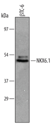
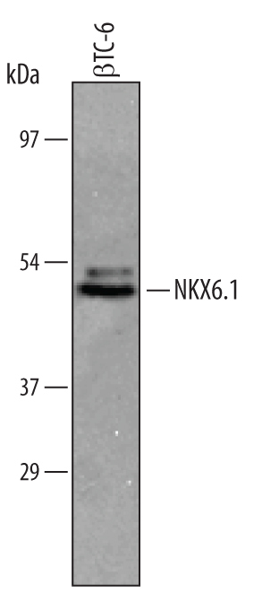
- Detection of Mouse NKX6.1 by Western Blot. Western blot shows lysates of beta TC-6 mouse beta cell insulinoma cell line. PVDF membrane was probed with 1 µg/mL of Goat Anti-Human/Mouse NKX6.1 Antigen Affinity-purified Polyclonal Antibody (Catalog # AF5857) followed by HRP-conjugated Anti-Goat IgG Secondary Antibody (Catalog # HAF019). A specific band was detected for NKX6.1 at approximately 46 kDa (as indicated). This experiment was conducted under reducing conditions and using Immunoblot Buffer Group 8.
- Submitted by
- R&D Systems (provider)
- Main image
- Experimental details
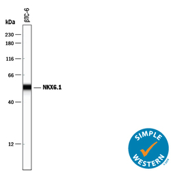
- Detection of Mouse NKX6.1 by Simple WesternTM. Simple Western lane view shows lysates of beta TC-6 mouse beta cell insulinoma cell line, loaded at 0.2 mg/mL. A specific band was detected for NKX6.1 at approximately 54 kDa (as indicated) using 10 µg/mL of Goat Anti-Human/Mouse NKX6.1 Antigen Affinity-purified Polyclonal Antibody (Catalog # AF5857) followed by 1:50 dilution of HRP-conjugated Anti-Goat IgG Secondary Antibody (Catalog # HAF109). This experiment was conducted under reducing conditions and using the 12-230 kDa separation system.

Supportive validation
- Submitted by
- R&D Systems (provider)
- Main image
- Experimental details
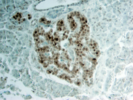
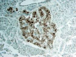
- NKX6.1 in Human Pancreas. NKX6.1 was detected in immersion fixed paraffin-embedded sections of human pancreas using Goat Anti-Human/Mouse NKX6.1 Antigen Affinity-purified Polyclonal Antibody (Catalog # AF5857) at 15 µg/mL overnight at 4 °C. Tissue was stained using the Anti-Goat HRP-DAB Cell & Tissue Staining Kit (brown; Catalog # CTS008) and counterstained with hematoxylin (blue). View our protocol for Chromogenic IHC Staining of Paraffin-embedded Tissue Sections.