 Explore
 Validate
 Learn
Learn Western blot
Antibody data
- Antibody Data
- Antigen structure
- References [2]
- Comments [0]
- Validations
- Western blot [6]
- Immunocytochemistry [1]
- Immunohistochemistry [1]
- Other assay [2]
Submit
Validation data
Reference
Comment
Report error
- Product number
- PA5-41442 - Provider product page

- Provider
- Invitrogen Antibodies
- Product name
- SOX4 Polyclonal Antibody
- Antibody type
- Polyclonal
- Antigen
- Synthetic peptide
- Description
- Peptide sequence: STASTGGKAD DPSWCKTPSG HIKRPMNAFM VWSQIERRKI MEQSPDMHNA
- Concentration
- 0.5 mg/mL
Submitted references LncRNA SNHG17 regulates cell proliferation and invasion by targeting miR-338-3p/SOX4 axis in esophageal squamous cell carcinoma.
MiR-34c downregulation leads to SOX4 overexpression and cisplatin resistance in nasopharyngeal carcinoma.
Chen W, Wang L, Li X, Zhao C, Shi L, Zhao H, Huang C
Cell death & disease 2021 Aug 24;12(9):806
Cell death & disease 2021 Aug 24;12(9):806
MiR-34c downregulation leads to SOX4 overexpression and cisplatin resistance in nasopharyngeal carcinoma.
Bissey PA, Teng M, Law JH, Shi W, Bruce JP, Petit V, Tsao SW, Yip KW, Liu FF
BMC cancer 2020 Jun 26;20(1):597
BMC cancer 2020 Jun 26;20(1):597
No comments: Submit comment
Supportive validation
- Submitted by
- Invitrogen Antibodies (provider)
- Main image

- Experimental details
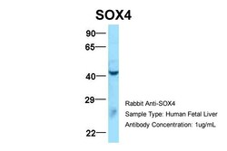
- Western blot analysis of human fetal liver cells using an anti-SOX4 polyclonal antibody (Product # PA5-41442).

- Submitted by
- Invitrogen Antibodies (provider)
- Main image

- Experimental details
- Western blot was performed using Anti-SOX4 Polyclonal Antibody (Product # PA5-41442) and a 47 kDa band corresponding to Transcription factor SOX-4 was observed across all the three lanes. Whole cell extracts (30 µg lysate) of Jurkat (Lane 1), Reh (Lane 2), U-2 OS (Lane 3) were electrophoresed using NuPAGE™ 4-12% Bis-Tris Protein Gel (Product # NP0322BOX). Resolved proteins were then transferred onto a nitrocellulose membrane (Product # IB23001) by iBlot® 2 Dry Blotting System (Product # IB21001). The blot was probed with the primary antibody (1:1000) and detected by chemiluminescence with Goat anti-Rabbit IgG (H+L) Superclonal™ Recombinant Secondary Antibody, HRP (Product # A27036,1:10000) using the iBright FL 1000 (Product # A32752). Chemiluminescentdetection was performed using Novex® ECL Chemiluminescent Substrate Reagent Kit (Product # WP20005).

- Submitted by
- Invitrogen Antibodies (provider)
- Main image
- Experimental details
- Western blot analysis of human fetal liver cells using an anti-SOX4 polyclonal antibody (Product # PA5-41442).

- Submitted by
- Invitrogen Antibodies (provider)
- Main image
- Experimental details
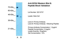
- Western blot analysis of human Hela cell lysate using an anti-SOX4 polyclonal antibody (Product # PA5-41442). Lane 1) Primary Antibody; Lane 2) Primary Antibody + Blocking Peptide.

- Submitted by
- Invitrogen Antibodies (provider)
- Main image

- Experimental details
- Western blot analysis of human HeLa cell lysate using an anti-SOX4 polyclonal antibody (Product # PA5-41442).

- Submitted by
- Invitrogen Antibodies (provider)
- Main image

- Experimental details
- Western blot analysis of human SH-SY5Y-RA cells using an anti-SOX4 polyclonal antibody (Product # PA5-41442).

Supportive validation
- Submitted by
- Invitrogen Antibodies (provider)
- Main image

- Experimental details
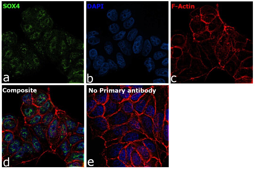
- Immunofluorescence analysis of Transcription factor SOX-4 was performed using 70% confluent log phase MCF7 cells. The cells were fixed with 4% paraformaldehyde for 10 minutes, permeabilized with 0.1% Triton™ X-100 for 15 minutes, and blocked with 2% BSA for 45 minutes at room temperature. The cells were labeled with SOX4 Polyclonal Antibody (Product # PA5-41442) at 1:200 in 0.1% BSA, incubated at 4 degree celsius overnight and then labeled with Goat anti-Rabbit IgG (H+L) Superclonal™ Recombinant Secondary Antibody, Alexa Fluor® 488 conjugate (Product # A27034), (1:2500), for 45 minutes at room temperature (Panel a: Green). Nuclei (Panel b:Blue) were stained with ProLong™ Diamond Antifade Mountant with DAPI (Product # P36962). F-actin (Panel c: Red) was stained with Rhodamine Phalloidin (Product # R415, 1:300). Panel d represents the merged image showing Primarily nuclear localization. Panel e represents control cells with no primary antibody to assess background. The images were captured at 60X magnification.
Supportive validation
- Submitted by
- Invitrogen Antibodies (provider)
- Main image

- Experimental details
- Immunohistochemistry analysis of human testis tissue using an anti-SOX4 polyclonal antibody (Product # PA5-41442).

Supportive validation
- Submitted by
- Invitrogen Antibodies (provider)
- Main image

- Experimental details
- Fig. 6 miR-338-3p directly targets SOX4 mRNA in ESCC cells. A The bioinformatics analysis of miRNA database of miR-338-3p. B Relative luciferase activity is decreased in cells transfected with pGL3-SOX4-WT1/2 and miR-338 mimic than in cells transfected with SOX4-MT1/2 and miR-338 mimic. * P < 0.05 vs. NC. C The data from starBase showed that SOX4 expression was upregulated in ESCC tissues compared to normal tissues. D , E The SOX4 expression was detected by RT-qPCR and IHC in clinical samples. * P < 0.05 vs. control group. F , G The RT-qPCR and western blot results showed that SOX4 expression level was increased by a miR-338 mimic in ESCC cells. H , I SNHG17 downregulation reduced the level of SOX4, and this was reversed by miR-338 inhibitor in cells. * P < 0.05 vs. NC.

- Submitted by
- Invitrogen Antibodies (provider)
- Main image

- Experimental details
- Fig. 7 Restoration of SOX4 reverses the effects of miR-338-3p on ESCC cells proliferation, invasion, and EMT process. A - C CCK-8, colony formation assays, and EdU analysis were performed to determine the proliferation ability of ESCC cells co-transfected with miR-338 mimic and SOX4, respectively. D - F Wound healing and transwell assays and western blot showed the invasion ability and EMT phenotype in ESCC cells co-transfected with miR-338 mimic and SOX4. * P < 0.05 vs. NC.
