Explore
 Validate
 Learn
Learn Western blot
Antibody data
- Antibody Data
- Antigen structure
- References [1]
- Comments [0]
- Validations
- Western blot [1]
- Immunohistochemistry [5]
- Other assay [1]
Submit
Validation data
Reference
Comment
Report error
- Product number
- PA5-27222 - Provider product page

- Provider
- Invitrogen Antibodies
- Product name
- AGR3 Polyclonal Antibody
- Antibody type
- Polyclonal
- Antigen
- Recombinant protein fragment
- Description
- Recommended positive controls: A549, MCF-7.
- Concentration
- 0.8 mg/mL
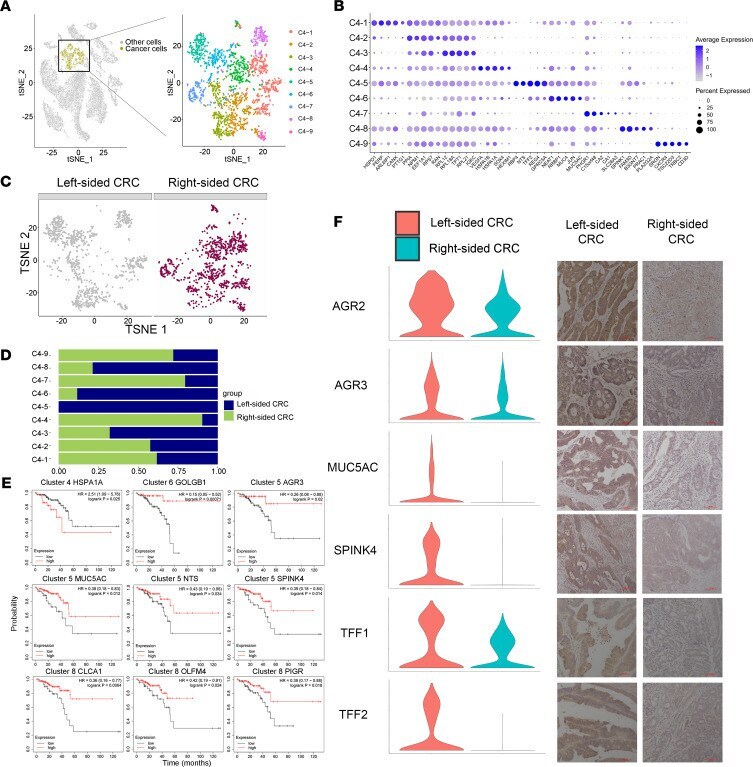
Submitted references Resolving the difference between left-sided and right-sided colorectal cancer by single-cell sequencing.
Guo W, Zhang C, Wang X, Dou D, Chen D, Li J
JCI insight 2022 Jan 11;7(1)
JCI insight 2022 Jan 11;7(1)
No comments: Submit comment
Supportive validation
- Submitted by
- Invitrogen Antibodies (provider)
- Main image

- Experimental details
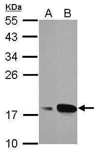
- Western Blot using AGR3 Polyclonal Antibody (Product # PA5-27222). Sample (30 µg of whole cell lysate). Lane A: A549. Lane B: MCF-7. 12% SDS PAGE. AGR3 Polyclonal Antibody (Product # PA5-27222) diluted at 1:1,000.
Supportive validation
- Submitted by
- Invitrogen Antibodies (provider)
- Main image

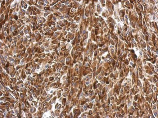
- Experimental details
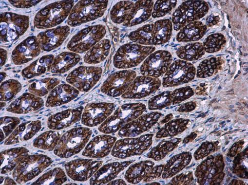
- AGR3 Polyclonal Antibody detects AGR3 protein at cytoplasm in rat colon by immunohistochemical analysis. Sample: Paraffin-embedded rat colon. AGR3 Polyclonal Antibody (Product # PA5-27222) diluted at 1:500. Antigen Retrieval: Citrate buffer, pH 6.0, 15 min.
- Submitted by
- Invitrogen Antibodies (provider)
- Main image

- Experimental details
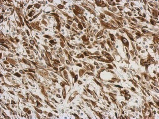
- Immunohistochemical analysis of paraffin-embedded C2C12 xenograft, using AGR3 (Product # PA5-27222) antibody at 1:500 dilution. Antigen Retrieval: EDTA based buffer, pH 8.0, 15 min.
- Submitted by
- Invitrogen Antibodies (provider)
- Main image
- Experimental details
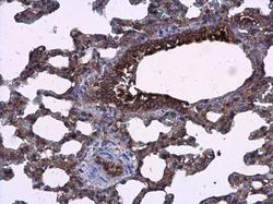
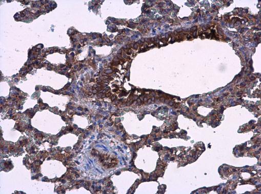
- AGR3 Polyclonal Antibody detects AGR3 protein at cytoplasm in rat lung by immunohistochemical analysis. Sample: Paraffin-embedded rat lung. AGR3 Polyclonal Antibody (Product # PA5-27222) diluted at 1:500. Antigen Retrieval: Citrate buffer, pH 6.0, 15 min.
- Submitted by
- Invitrogen Antibodies (provider)
- Main image

- Experimental details
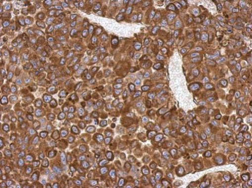
- Immunohistochemical analysis of paraffin-embedded PC13 xenograft, using AGR3 (Product # PA5-27222) antibody at 1:500 dilution. Antigen Retrieval: EDTA based buffer, pH 8.0, 15 min.
- Submitted by
- Invitrogen Antibodies (provider)
- Main image
- Experimental details
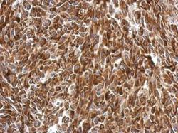
- Immunohistochemical analysis of paraffin-embedded RT2 xenograft, using AGR3 (Product # PA5-27222) antibody at 1:500 dilution. Antigen Retrieval: EDTA based buffer, pH 8.0, 15 min.
Supportive validation
- Submitted by
- Invitrogen Antibodies (provider)
- Main image

- Experimental details
- Figure 7 A RBP4 + NTS + cancer cell subset is unique to left-sided CRC. ( A ) The t-SNE plot that showed the distribution of cancer cell lineages (yellow, n = 2196 cells) within the atlas. Cancer cell populations were reclustered into 9 subclusters (color coding). ( B ) Top 5 marker genes of 15 major cell types identified in this profile. ( C ) Annotation by left-sided and right-sided CRC cells. ( D ) The fraction of cells that originated from left-sided and right-sided CRC samples for 9 subgroups identified in this profile. ( E ) Kaplan-Meier survival curves of OS based on HSPA1A, GOLGB1, AGR3, MUC5AC, NTS, SPINK4, CLCA1, OLFM4, and PIGR expression using the online bioinformatics tool Kaplan-Meier Plotter. ( F ) Violin plots and immunochemistry display the distribution of expression of AGR2, AGR3, MUC5AC, SPINK4, TFF1, and TFF2 between left-sided and right-sided CRC. Scale bars: 100 um.