Explore
 Validate
 Learn
Learn Western blot
 Immunohistochemistry
Antibody data
- Antibody Data
- Antigen structure
- References [1]
- Comments [0]
- Validations
- Immunohistochemistry [8]
Submit
Validation data
Reference
Comment
Report error
- Product number
- NB100-2737 - Provider product page

- Provider
- Novus Biologicals
- Proper citation
- Novus Cat#NB100-2737, RRID:AB_10003423
- Product name
- Mouse Monoclonal PD-ECGF/Thymidine Phosphorylase Antibody
- Antibody type
- Monoclonal
- Description
- Protein G purified.
- Reactivity
- Human
- Host
- Mouse
- Isotype
- IgG
- Vial size
- 0.1 mg
- Concentration
- 1 mg/ml
- Storage
- Store at 4C. Do not freeze.
Submitted references Expression of thymidine phosphorylase in lymph nodes involved with mycosis fungoides and sézary syndrome.
Nie X, Bhat R, Al-Saleem ED, Vonderheid EC, Hou JS
Advances in hematology 2011;2011:875135
Advances in hematology 2011;2011:875135
No comments: Submit comment
Supportive validation
- Submitted by
- Novus Biologicals (provider)
- Main image
- Experimental details
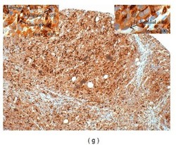
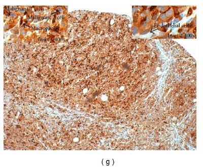
- Immunohistochemistry: PD-ECGF/Thymidine Phosphorylase Antibody (PGF 44C) [NB100-2737] - Immunohistochemical staining of thymidine phosphorylase (TP) expression and related markers in benign lymph node in lymph node with malignant mycosis fungoides/Sezary syndrome cells (LN-MF). TP highlights the macrophages (cytoplasmic/nuclear pattern, arrowhead in inset 1; 400x) and the malignant T cells (cytoplasmic pattern, arrow in inset 2; 400x) in a cytoplasmic staining pattern. Multiple mitotic figures are also noted in the malignant T cells. Images are from an Olympus BX41 microscope (Olympus Corp., Tokyo, Japan) processed with Qcapture Pro 5.1 (QImaging, Surrey, BC, Canada). Image collected and cropped by CiteAb from the following publication (http://www.hindawi.com/journals/ah/2011/875135/) licensed under a CC-BY licence.
- Submitted by
- Novus Biologicals (provider)
- Main image
- Experimental details
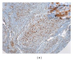
- Immunohistochemistry: PD-ECGF/Thymidine Phosphorylase Antibody (PGF 44C) [NB100-2737] - Immunohistochemical staining of thymidine phosphorylase (TP) expression and related markers in benign lymph node. In benign lymph node, TP expression highlights the meshwork of macrophages (arrow in inset; 400x) and endothelial cells (arrowhead in inset; 400x). Images are from an Olympus BX41 microscope (Olympus Corp., Tokyo, Japan) processed with Qcapture Pro 5.1 (QImaging, Surrey, BC, Canada). Image collected and cropped by CiteAb from the following publication (http://www.hindawi.com/journals/ah/2011/875135/) licensed under a CC-BY licence.

- Submitted by
- Novus Biologicals (provider)
- Main image
- Experimental details
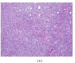
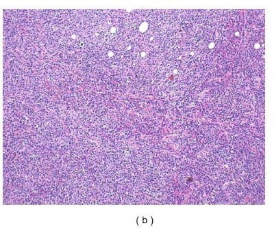
- Immunohistochemistry: PD-ECGF/Thymidine Phosphorylase Antibody (PGF 44C) [NB100-2737] - Immunohistochemical staining of thymidine phosphorylase (TP) expression and related markers in benign lymph node in lymph node with malignant mycosis fungoides/Sezary syndrome cells (LN-MF). LN-MF: hematoxylin and eosin (H&E) stain. Images are from an Olympus BX41 microscope (Olympus Corp., Tokyo, Japan) processed with Qcapture Pro 5.1 (QImaging, Surrey, BC, Canada). Image collected and cropped by CiteAb from the following publication (http://www.hindawi.com/journals/ah/2011/875135/) licensed under a CC-BY licence.
- Submitted by
- Novus Biologicals (provider)
- Main image
- Experimental details
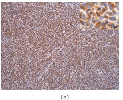
- Immunohistochemistry: PD-ECGF/Thymidine Phosphorylase Antibody (PGF 44C) [NB100-2737] - Immunohistochemical staining of thymidine phosphorylase (TP) expression and related markers in benign lymph node in lymph node with malignant mycosis fungoides/Sezary syndrome cells (LN-MF). CD3 highlights the malignant T cells and reactive T cells in LN-MF. Images are from an Olympus BX41 microscope (Olympus Corp., Tokyo, Japan) processed with Qcapture Pro 5.1 (QImaging, Surrey, BC, Canada). Image collected and cropped by CiteAb from the following publication (http://www.hindawi.com/journals/ah/2011/875135/) licensed under a CC-BY licence.

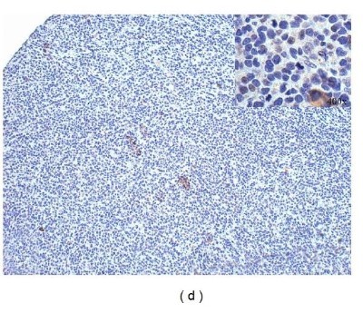
- Submitted by
- Novus Biologicals (provider)
- Main image
- Experimental details
- Immunohistochemistry: PD-ECGF/Thymidine Phosphorylase Antibody (PGF 44C) [NB100-2737] - Immunohistochemical staining of thymidine phosphorylase (TP) expression and related markers in benign lymph node in lymph node with malignant mycosis fungoides/Sezary syndrome cells (LN-MF). CD4 is weakly expressed in the malignant T cells in LN-MF. Images are from an Olympus BX41 microscope (Olympus Corp., Tokyo, Japan) processed with Qcapture Pro 5.1 (QImaging, Surrey, BC, Canada). Image collected and cropped by CiteAb from the following publication (http://www.hindawi.com/journals/ah/2011/875135/) licensed under a CC-BY licence.
- Submitted by
- Novus Biologicals (provider)
- Main image
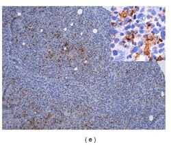
- Experimental details
- Immunohistochemistry: PD-ECGF/Thymidine Phosphorylase Antibody (PGF 44C) [NB100-2737] - Immunohistochemical staining of thymidine phosphorylase (TP) expression and related markers in benign lymph node in lymph node with malignant mycosis fungoides/Sezary syndrome cells (LN-MF). CD68 highlights the macrophages in LN-MF. Images are from an Olympus BX41 microscope (Olympus Corp., Tokyo, Japan) processed with Qcapture Pro 5.1 (QImaging, Surrey, BC, Canada). Image collected and cropped by CiteAb from the following publication (http://www.hindawi.com/journals/ah/2011/875135/) licensed under a CC-BY licence.

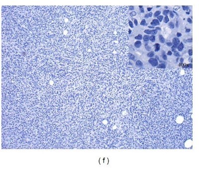
- Submitted by
- Novus Biologicals (provider)
- Main image

- Experimental details
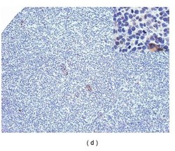
- Immunohistochemistry: PD-ECGF/Thymidine Phosphorylase Antibody (PGF 44C) [NB100-2737] - Immunohistochemical staining of thymidine phosphorylase (TP) expression and related markers in benign lymph node in lymph node with malignant mycosis fungoides/Sezary syndrome cells (LN-MF). CD21 is negative in this case of LN-MF. Images are from an Olympus BX41 microscope (Olympus Corp., Tokyo, Japan) processed with Qcapture Pro 5.1 (QImaging, Surrey, BC, Canada). Image collected and cropped by CiteAb from the following publication (http://www.hindawi.com/journals/ah/2011/875135/) licensed under a CC-BY licence.
- Submitted by
- Novus Biologicals (provider)
- Main image

- Experimental details
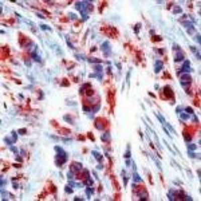
- Immunohistochemistry-Paraffin: PD-ECGF/Thymidine Phosphorylase Antibody (PGF 44C) [NB100-2737] - Analysis of Thymidine Phosphorylase formalin-fixed paraffin embedded human breast carcinoma.