Explore
 Validate
 Learn
Learn Western blot
Antibody data
- Antibody Data
- Antigen structure
- References [0]
- Comments [0]
- Validations
- Western blot [3]
- Immunocytochemistry [1]
- Immunohistochemistry [6]
Submit
Validation data
Reference
Comment
Report error
- Product number
- HPA030646 - Provider product page

- Provider
- Atlas Antibodies
- Proper citation
- Atlas Antibodies Cat#HPA030646, RRID:AB_10602019
- Product name
- Anti-PGGT1B
- Antibody type
- Polyclonal
- Reactivity
- Human, Mouse, Rat
- Host
- Rabbit
- Conjugate
- Unconjugated
- Antigen sequence
GAGLESHGGSTFCGIASLCLMGKLEEVFSEKELNR
IKRWCIMRQQNGYHGRPNKPVDTCYSFWVGATLKL
LKIFQYTNFEKNRNYILSTQDRLVGGFAKWPDSHP
DALHAYFGICGLSLMEESGICKVHPALNVS- Isotype
- IgG
- Vial size
- 100 µl
- Storage
- Store at +4°C for short term storage. Long time storage is recommended at -20°C.
No comments: Submit comment
Supportive validation
- Submitted by
- Atlas Antibodies (provider)
- Main image

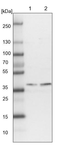
- Experimental details
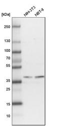
- Lane 1: NIH-3T3 cell lysate (Mouse embryonic fibroblast cells)Lane 2: NBT-II cell lysate (Rat Wistar bladder tumour cells)
- Submitted by
- Atlas Antibodies (provider)
- Main image
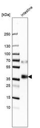
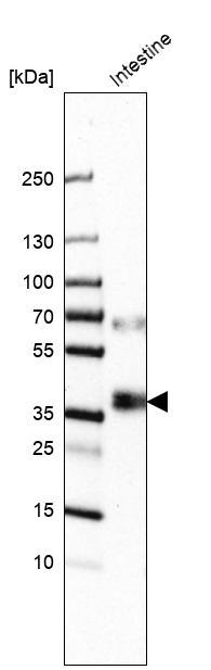
- Experimental details
- Western blot analysis in human small intestine tissue.
- Submitted by
- Atlas Antibodies (provider)
- Main image
- Experimental details
- Western blot analysis in mouse cell line NIH-3T3 and rat cell line NBT-II.

Supportive validation
- Submitted by
- Atlas Antibodies (provider)
- Main image

- Experimental details
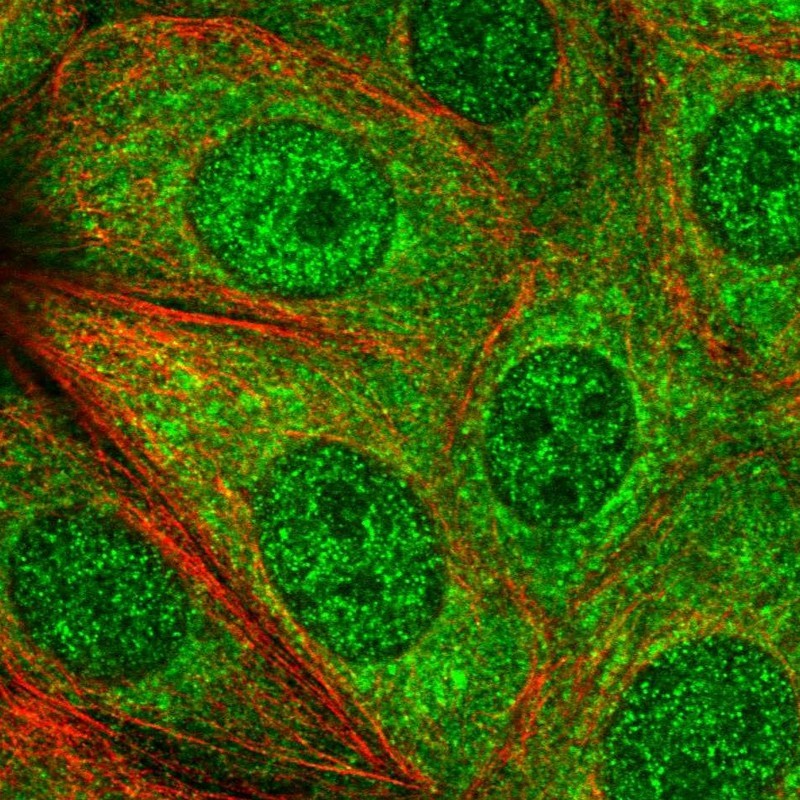
- Immunofluorescent staining of human cell line MCF7 shows positivity in cytoplasm & nucleus but excluded from the nucleoli.
- Sample type
- HUMAN
Supportive validation
- Submitted by
- Atlas Antibodies (provider)
- Main image

- Experimental details
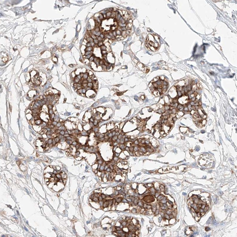
- Immunohistochemical staining of human breast shows distinct cytoplasmic positivity in glandular cells.
- Submitted by
- Atlas Antibodies (provider)
- Main image
- Experimental details
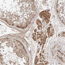
- Immunohistochemical staining of human testis shows moderate cytoplasmic positivity in Leydig cells.
- Sample type
- HUMAN

- Submitted by
- Atlas Antibodies (provider)
- Main image
- Experimental details
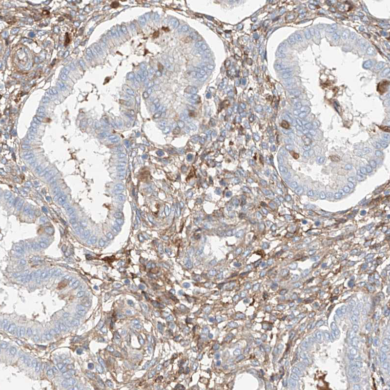
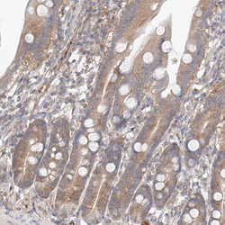
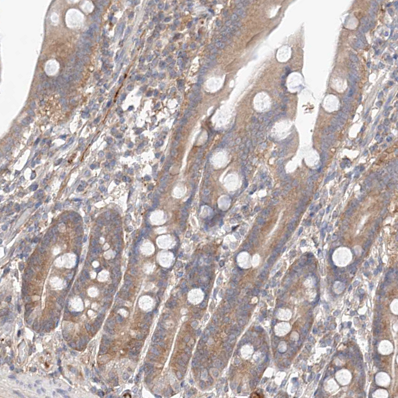
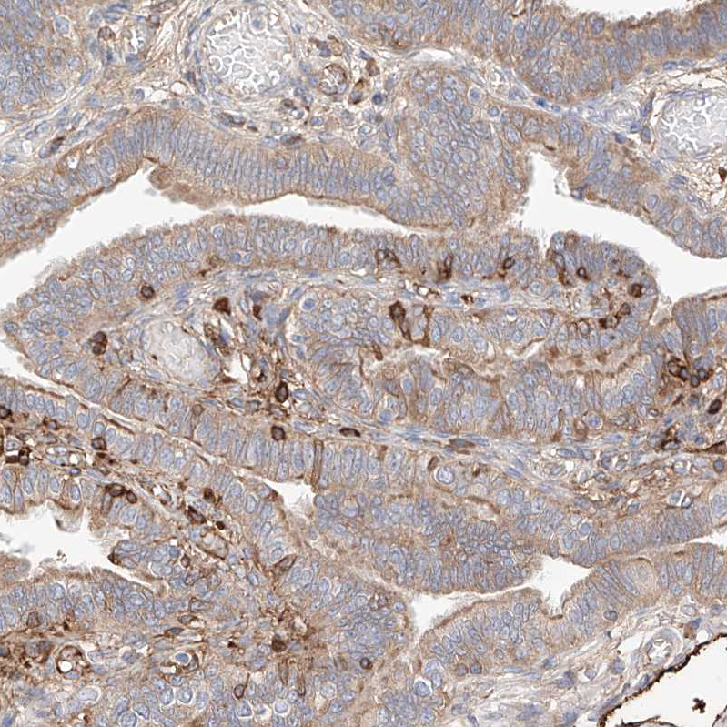
- Immunohistochemical staining of human duodenum shows moderate tcytoplasmic positivity in glandular cells.
- Sample type
- HUMAN
- Submitted by
- Atlas Antibodies (provider)
- Main image
- Experimental details
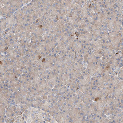
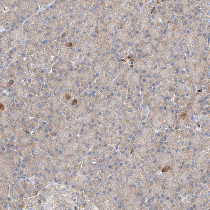
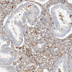
- Immunohistochemical staining of human pancreas shows weak cytoplasmic positivity in exocrine glandular cells.
- Sample type
- HUMAN
- Submitted by
- Atlas Antibodies (provider)
- Main image

- Experimental details
- Immunohistochemical staining of human Fallopian tube shows strong cytoplasmic positivity in stromal cells.
- Sample type
- HUMAN
- Submitted by
- Atlas Antibodies (provider)
- Main image
- Experimental details
- Immunohistochemical staining of human endometrium shows moderate cytoplasmic positivity in stromal cells.
- Sample type
- HUMAN