 Explore
 Validate
 Learn
Learn51-5600
antibody from Invitrogen Antibodies
Targeting: LIN7C
FLJ11215, LIN-7-C, LIN-7C, MALS-3, VELI3
 Western blot
 ELISA Immunocytochemistry Immunoprecipitation Immunohistochemistry Flow cytometry Other assay
Antibody data
- Antibody Data
- Antigen structure
- References [12]
- Comments [0]
- Validations
- Western blot [1]
- Immunocytochemistry [1]
- Flow cytometry [1]
- Other assay [2]
Submit
Validation data
Reference
Comment
Report error
- Product number
- 51-5600 - Provider product page

- Provider
- Invitrogen Antibodies
- Product name
- LIN7C Polyclonal Antibody
- Antibody type
- Polyclonal
- Antigen
- Synthetic peptide
- Reactivity
- Human, Mouse, Rat, Canine
- Host
- Rabbit
- Isotype
- IgG
- Vial size
- 100 µg
- Concentration
- 0.25 mg/mL
- Storage
- -20° C, Avoid Freeze/Thaw Cycles
Submitted references Calcium/calmodulin-dependent serine protein kinase (CASK), a protein implicated in mental retardation and autism-spectrum disorders, interacts with T-Brain-1 (TBR1) to control extinction of associative memory in male mice.
NHERF1 in Microvilli of Vomeronasal Sensory Neurons.
Defective ceramide synthases in mice cause reduced amplitudes in electroretinograms and altered sphingolipid composition in retina and cornea.
Expression and Localization of Connexins in the Outer Retina of the Mouse.
Evidence for a Clathrin-independent mode of endocytosis at a continuously active sensory synapse.
Testing for a gap junction-mediated bystander effect in retinitis pigmentosa: secondary cone death is not altered by deletion of connexin36 from cones.
miR-199a-5p regulates urothelial permeability and may play a role in bladder pain syndrome.
Stability of active zone components at the photoreceptor ribbon complex.
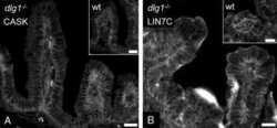
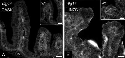
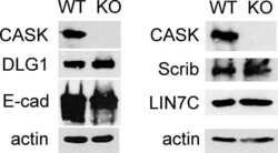
CASK deletion in intestinal epithelia causes mislocalization of LIN7C and the DLG1/Scrib polarity complex without affecting cell polarity.
Type 4 OFF cone bipolar cells of the mouse retina express calsenilin and contact cones as well as rods.
The serotonin 5-HT2A and 5-HT2C receptors interact with specific sets of PDZ proteins.
Synaptic multiprotein complexes associated with 5-HT(2C) receptors: a proteomic approach.
Huang TN, Hsueh YP
Journal of psychiatry & neuroscience : JPN 2017 Jan;42(1):37-47
Journal of psychiatry & neuroscience : JPN 2017 Jan;42(1):37-47
NHERF1 in Microvilli of Vomeronasal Sensory Neurons.
Henkel B, Bintig W, Bhat SS, Spehr M, Neuhaus EM
Chemical senses 2017 Jan;42(1):25-35
Chemical senses 2017 Jan;42(1):25-35
Defective ceramide synthases in mice cause reduced amplitudes in electroretinograms and altered sphingolipid composition in retina and cornea.
Brüggen B, Kremser C, Bickert A, Ebel P, Vom Dorp K, Schultz K, Dörmann P, Willecke K, Dedek K
The European journal of neuroscience 2016 Jul;44(1):1700-13
The European journal of neuroscience 2016 Jul;44(1):1700-13
Expression and Localization of Connexins in the Outer Retina of the Mouse.
Bolte P, Herrling R, Dorgau B, Schultz K, Feigenspan A, Weiler R, Dedek K, Janssen-Bienhold U
Journal of molecular neuroscience : MN 2016 Feb;58(2):178-92
Journal of molecular neuroscience : MN 2016 Feb;58(2):178-92
Evidence for a Clathrin-independent mode of endocytosis at a continuously active sensory synapse.
Fuchs M, Brandstätter JH, Regus-Leidig H
Frontiers in cellular neuroscience 2014;8:60
Frontiers in cellular neuroscience 2014;8:60
Testing for a gap junction-mediated bystander effect in retinitis pigmentosa: secondary cone death is not altered by deletion of connexin36 from cones.
Kranz K, Paquet-Durand F, Weiler R, Janssen-Bienhold U, Dedek K
PloS one 2013;8(2):e57163
PloS one 2013;8(2):e57163
miR-199a-5p regulates urothelial permeability and may play a role in bladder pain syndrome.
Monastyrskaya K, Sánchez-Freire V, Hashemi Gheinani A, Klumpp DJ, Babiychuk EB, Draeger A, Burkhard FC
The American journal of pathology 2013 Feb;182(2):431-48
The American journal of pathology 2013 Feb;182(2):431-48
Stability of active zone components at the photoreceptor ribbon complex.
Regus-Leidig H, Specht D, Tom Dieck S, Brandstätter JH
Molecular vision 2010 Dec 12;16:2690-700
Molecular vision 2010 Dec 12;16:2690-700
CASK deletion in intestinal epithelia causes mislocalization of LIN7C and the DLG1/Scrib polarity complex without affecting cell polarity.
Lozovatsky L, Abayasekara N, Piawah S, Walther Z
Molecular biology of the cell 2009 Nov;20(21):4489-99
Molecular biology of the cell 2009 Nov;20(21):4489-99
Type 4 OFF cone bipolar cells of the mouse retina express calsenilin and contact cones as well as rods.
Haverkamp S, Specht D, Majumdar S, Zaidi NF, Brandstätter JH, Wasco W, Wässle H, Tom Dieck S
The Journal of comparative neurology 2008 Mar 1;507(1):1087-101
The Journal of comparative neurology 2008 Mar 1;507(1):1087-101
The serotonin 5-HT2A and 5-HT2C receptors interact with specific sets of PDZ proteins.
Bécamel C, Gavarini S, Chanrion B, Alonso G, Galéotti N, Dumuis A, Bockaert J, Marin P
The Journal of biological chemistry 2004 May 7;279(19):20257-66
The Journal of biological chemistry 2004 May 7;279(19):20257-66
Synaptic multiprotein complexes associated with 5-HT(2C) receptors: a proteomic approach.
Bécamel C, Alonso G, Galéotti N, Demey E, Jouin P, Ullmer C, Dumuis A, Bockaert J, Marin P
The EMBO journal 2002 May 15;21(10):2332-42
The EMBO journal 2002 May 15;21(10):2332-42
No comments: Submit comment
Supportive validation
- Submitted by
- Invitrogen Antibodies (provider)
- Main image
- Experimental details
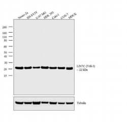
- Western blot analysis was performed on whole cell extracts (30 µg lysate) of Neuro-2a (Lane 1), SH-SY5Y (Lane 2), U-87 MG (Lane 3), HEK 293 (Lane 4), Caki-1 (Lane 5), COS-7 (Lane 6), and MDCK (Lane 7). The blots were probed with Rabbit Anti-Veli-3 Polyclonal Antibody (Product # 51-5600, 2 µg/mL) and detected by chemiluminescence using Goat anti-Rabbit IgG (H+L) Superclonal™ Secondary Antibody, HRP conjugate (Product # A27036, 0.25 µg/mL, 1:4000 dilution). A 22 kDa band corresponding to LIN7C (Veli-3) was observed across the cell lines tested. Known quantity of protein samples were electrophoresed using Novex® NuPAGE® 4-12 % Bis-Tris gel (Product # NP0321BOX), XCell SureLock™ Electrophoresis System (Product # EI0002) and Novex® Sharp Pre-Stained Protein Standard (Product # LC5800). Resolved proteins were then transferred onto a nitrocellulose membrane with iBlot® 2 Dry Blotting System (Product # IB21001). The membrane was probed with the relevant primary and secondary Antibody following blocking with 5 % skimmed milk. Chemiluminescent detection was performed using Pierce™ ECL Western Blotting Substrate (Product # 32106).

Supportive validation
- Submitted by
- Invitrogen Antibodies (provider)
- Main image
- Experimental details
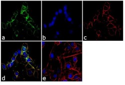
- Immunofluorescence analysis of VELI-3 was performed using 70% confluent log phase RSC96 cells. The cells were fixed with 4% paraformaldehyde for 10 minutes, permeabilized with 0.1% Triton™ X-100 for 10 minutes, and blocked with 1% BSA for 1 hour at room temperature. The cells were labeled with LIN7C Rabbit Polyclonal Antibody (Product # 51-5600) at 2µg/mL in 0.1% BSA and incubated for 3 hours at room temperature and then labeled with Goat anti-Rabbit IgG (H+L) Superclonal™ Secondary Antibody, Alexa Fluor® 488 conjugate (Product # A27034) at a dilution of 1:2000 for 45 minutes at room temperature (Panel a: green). Nuclei (Panel b: blue) were stained with SlowFade® Gold Antifade Mountant with DAPI (Product # S36938). F-actin (Panel c: red) was stained with Alexa Fluor® 555 Rhodamine Phalloidin (Product # R415, 1:300). Panel d represents the merged image showing membranous localization. Panel e shows the no primary antibody control. The images were captured at 60X magnification.

Supportive validation
- Submitted by
- Invitrogen Antibodies (provider)
- Main image

- Experimental details
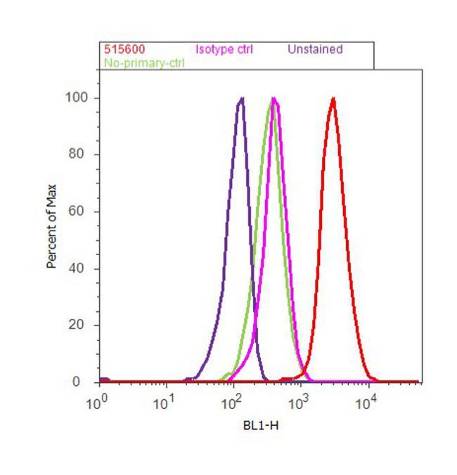
- Flow cytometry analysis of LIN7C was done on RSC96 cells. Cells were fixed with 70% ethanol for 10 minutes, permeabilized with 0.25% Triton™ X-100 for 20 minutes, and blocked with 5% BSA for 30 minutes at room temperature. Cells were labeled with LIN7C Rabbit Polyclonal Antibody (515600, red histogram) or with rabbit isotype control (pink histogram) at 3-5 ug/million cells in 2.5% BSA. After incubation at room temperature for 2 hours, the cells were labeled with Alexa Fluor® 488 Goat Anti-Rabbit Secondary Antibody (A11008) at a dilution of 1:400 for 30 minutes at room temperature. The representative 10,000 cells were acquired and analyzed for each sample using an Attune® Acoustic Focusing Cytometer. The purple histogram represents unstained control cells and the green histogram represents no-primary-antibody control..
Supportive validation
- Submitted by
- Invitrogen Antibodies (provider)
- Main image
- Experimental details
- NULL
- Submitted by
- Invitrogen Antibodies (provider)
- Main image
- Experimental details
- NULL
