Explore
 Validate
 Learn
Learn Western blot
 Immunohistochemistry
Antibody data
- Antibody Data
- Antigen structure
- References [0]
- Comments [0]
- Validations
- Western blot [1]
- Immunocytochemistry [5]
- Immunohistochemistry [4]
Submit
Validation data
Reference
Comment
Report error
- Product number
- AMAb90772 - Provider product page

- Provider
- Atlas Antibodies
- Proper citation
- Atlas Antibodies Cat#AMAb90772, RRID:AB_2665660
- Product name
- Anti-PLA2R1
- Antibody type
- Monoclonal
- Reactivity
- Human
- Host
- Mouse
- Conjugate
- Unconjugated
- Antigen sequence
EEKTWHEALRSCQADNSALIDITSLAEVEFLVTLL
GDENASETWIGLSSNKIPVSFEWSNDSSVIFTNWH
TLEPHIFPNRSQLCVSAEQSEGHWKVKNCEERLFY
ICKKAGHVLSDAESGCQEGWERHGGFCYKID- Epitope
- Binds to an epitope located within the peptide sequence KIPVSFEWSN as determined by overlapping synthetic peptides.
- Isotype
- IgG
- Antibody clone number
- CL0474
- Vial size
- 100 µl
- Storage
- Store at +4°C for short term storage. Long time storage is recommended at -20°C.
No comments: Submit comment
Supportive validation
- Submitted by
- Atlas Antibodies (provider)
- Main image
- Experimental details
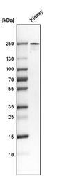
- Western blot analysis in human kidney tissue.

Supportive validation
- Submitted by
- Atlas Antibodies (provider)
- Main image

- Experimental details
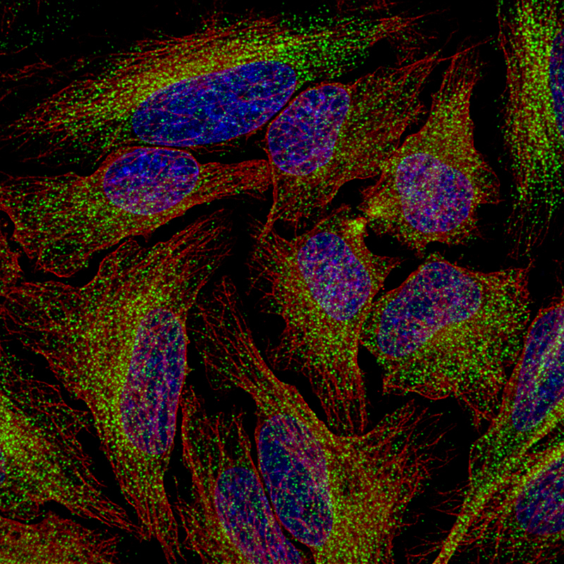
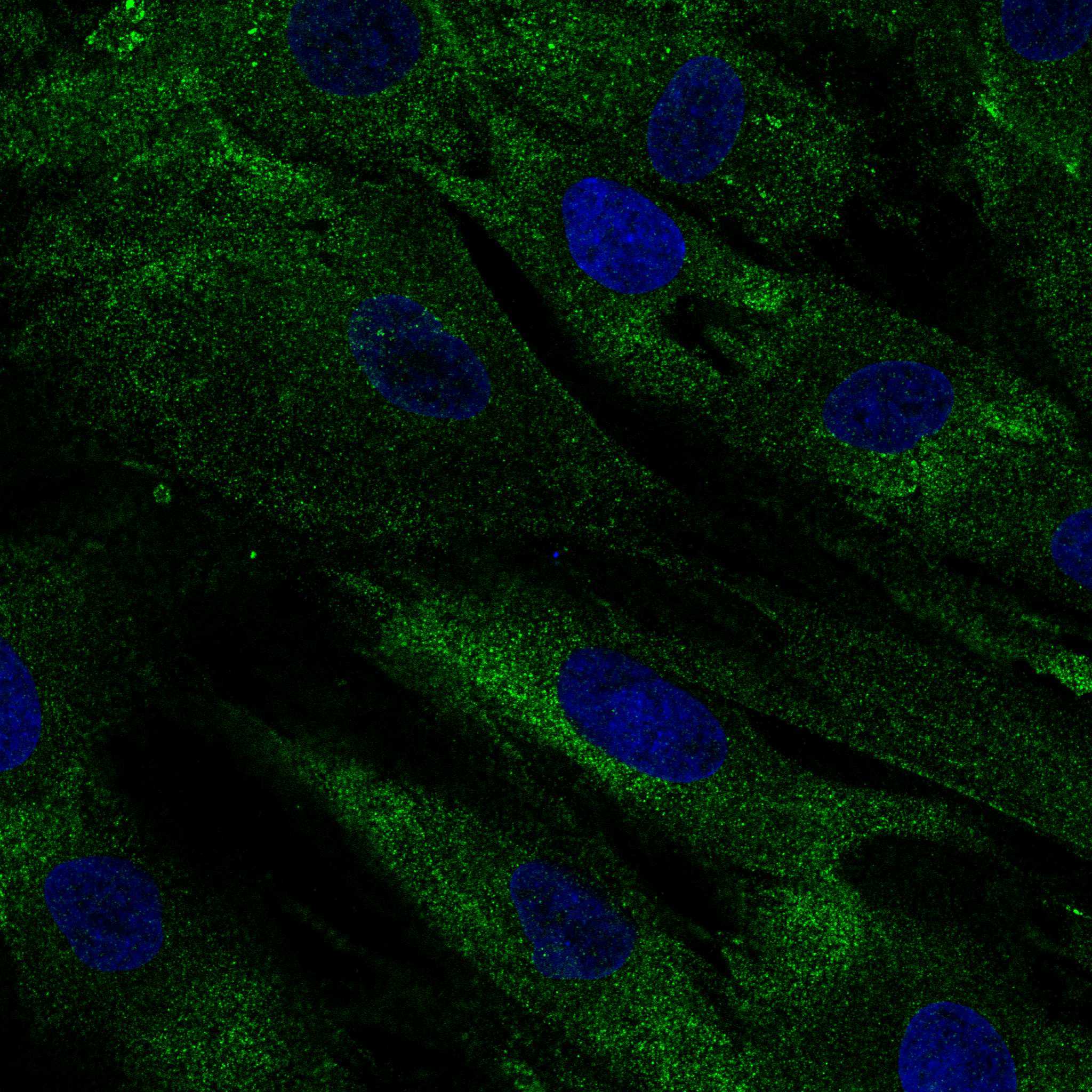
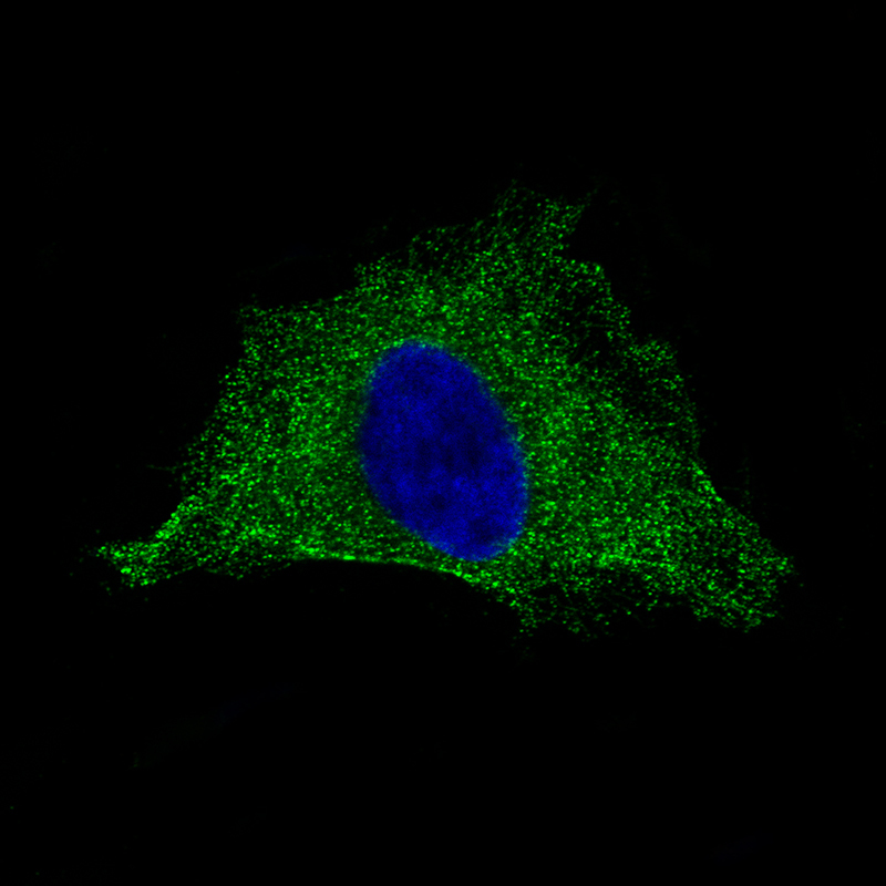
- Immunofluorescence staining of HeLa cells using the Anti-PLA2R1 monoclonal antibody, showing specific staining in the cytosol in green. Microtubule- and nuclear probes are visualized in red and blue, respectively (where available).
- Sample type
- HUMAN
- Submitted by
- Atlas Antibodies (provider)
- Main image
- Experimental details
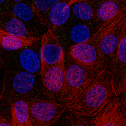
- Immunofluorescence staining of MCF7 cells using the Anti-PLA2R1 monoclonal antibody, showing only weak staining in cytosol. MCF7 cells serves as a negative control based on RNA-seq values. Microtubule- and nuclear probes are visualized in red and blue, respectively (where available).
- Sample type
- HUMAN

- Submitted by
- Atlas Antibodies (provider)
- Main image

- Experimental details
- Immunofluorescence staining of BJ cells using the anti-PLA2R1 monoclonal antibody, showing specific staining in the cytosol in green. Microtubule- and nuclear probes are visualized in red and blue, respectively (where available).
- Sample type
- HUMAN
- Submitted by
- Atlas Antibodies (provider)
- Main image

- Experimental details
- Immunofluorescence staining of HeLa cells using the Anti-PLA2R1 monoclonal antibody, showing specific staining in the cytosol and plasma membrane in green. Microtubule- and nuclear probes are visualized in red and blue, respectively (where available).
- Sample type
- HUMAN
- Submitted by
- Atlas Antibodies (provider)
- Main image
- Experimental details
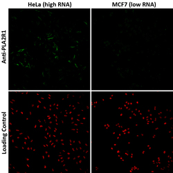
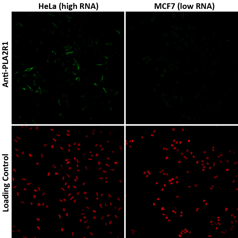
- Validation of the Anti-PLA2R1 monoclonal antibody by comparing the immunofluorescence staining in human cell lines Hela and MCF7 exhibiting a relatively high and low expression of PLA2R1 (based on RNA-seq values), respectively. The PLA2R1 (green signal) is present in Hela cells but is absent in MCF7 cells. The anti-HDAC1 antibody (HPA029693) was used as a loading control (red signal).
- Sample type
- HUMAN
Enhanced validation
Supportive validation
- Submitted by
- Atlas Antibodies (provider)
- Enhanced method
- Orthogonal validation
- Main image

- Experimental details
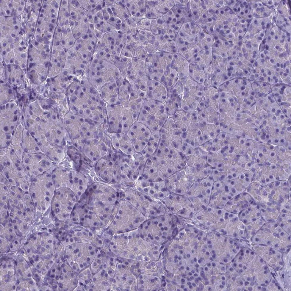
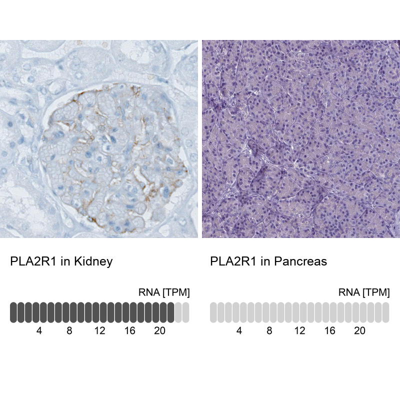
- Immunohistochemistry analysis in human kidney and pancreas tissues using AMAb90772 antibody. Corresponding PLA2R1 RNA-seq data are presented for the same tissues.
- Sample type
- HUMAN
Supportive validation
- Submitted by
- Atlas Antibodies (provider)
- Main image
- Experimental details
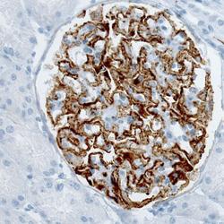
- Immunohistochemical staining of human kidney (idiopathic membranous nephropathy) shows strong membranous positivity in cells in glomeruli.

- Submitted by
- Atlas Antibodies (provider)
- Main image
- Experimental details
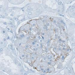
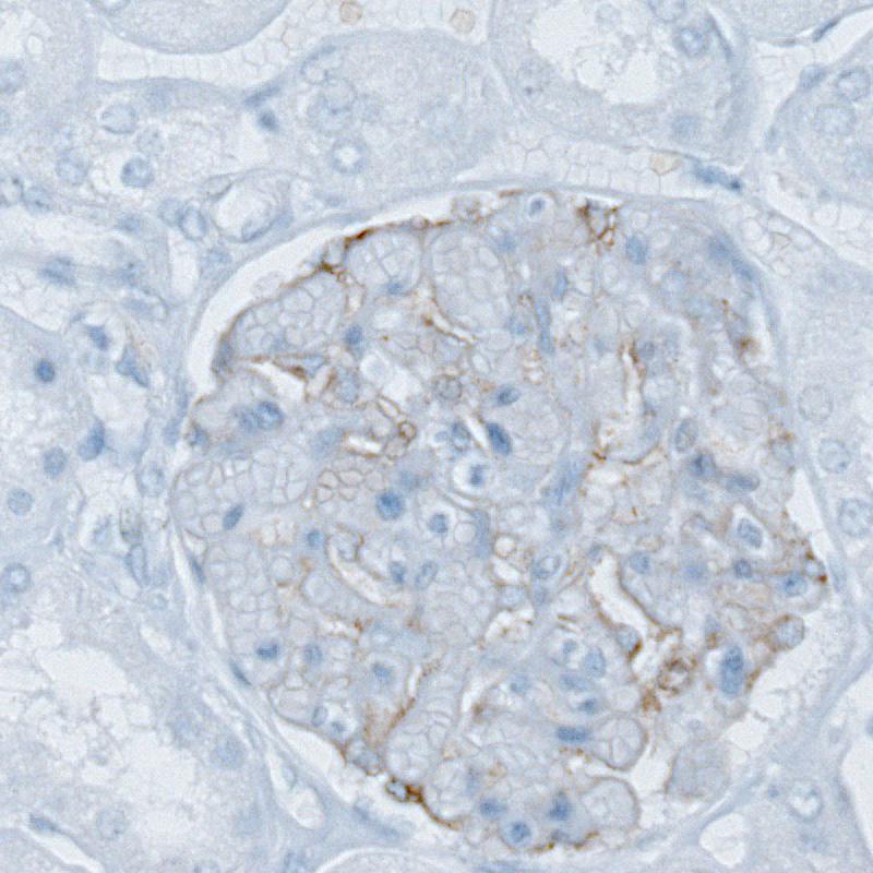
- Immunohistochemical staining of human kidney shows weak membranous positivity in cells in glomeruli.
- Submitted by
- Atlas Antibodies (provider)
- Main image

- Experimental details
- Immunohistochemical staining of human pancreas shows no positivity in exocrine glandular cells as expected.