Explore
 Validate
 Learn
Learn Western blot
 ELISA
ELISAAntibody data
- Antibody Data
- Antigen structure
- References [1]
- Comments [0]
- Validations
- Western blot [1]
- Immunohistochemistry [3]
- Flow cytometry [2]
Submit
Validation data
Reference
Comment
Report error
- Product number
- NBP2-50248 - Provider product page

- Provider
- Novus Biologicals
- Product name
- Mouse Monoclonal PLA2R1 Antibody
- Antibody type
- Monoclonal
Submitted references PLA2R binds to the annexin A2-S100A10 complex in human podocytes.
Fresquet M, Jowitt TA, McKenzie EA, Ball MD, Randles MJ, Lennon R, Brenchley PE
Scientific reports 2017 Jul 31;7(1):6876
Scientific reports 2017 Jul 31;7(1):6876
No comments: Submit comment
Supportive validation
- Submitted by
- Novus Biologicals (provider)
- Main image
- Experimental details
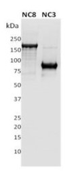
- Western Blot: PLA2R1 Antibody (12-6-5) [NBP2-50248] - 1ug of recombinant full length extracellular domain of human PLA2R (NC8) and a N-terminus fragment of PLA2R (NC3) were subjected to SDS PAGE followed by western blot using Ms anti-PLAR (Cl12-6-5) at dilution 1:10000.

Supportive validation
- Submitted by
- Novus Biologicals (provider)
- Main image
- Experimental details
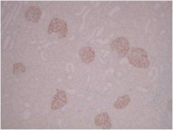
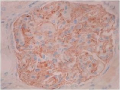
- Immunohistochemistry: PLA2R1 Antibody (12-6-5) [NBP2-50248] - Normal human kidney showing glomerular specific staining. IHC Protocol Info: HIER pH8 (CC1) for 64 minutes, 36 minute primary antibody (PLA2R Mouse 12-6-5) incubation at room temperature using a dilution of 1:5000, using Ventana Ultraview DAB detection system.
- Submitted by
- Novus Biologicals (provider)
- Main image

- Experimental details
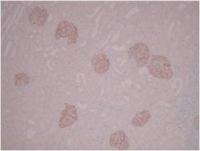
- Immunohistochemistry: PLA2R1 Antibody (12-6-5) [NBP2-50248] - Normal human kidney showing podocyte specific staining. IHC Protocol Info: HIER pH8 (CC1) for 64 minutes, 36 minute primary antibody (PLA2R Mouse 12-6-5) incubation at room temperature using a dilution of 1:5000, using Ventana Ultraview DAB detection system.
- Submitted by
- Novus Biologicals (provider)
- Main image

- Experimental details
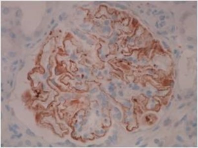
- Immunohistochemistry: PLA2R1 Antibody (12-6-5) [NBP2-50248] - Membranous nephropathy kidney showing staining of PLA2R in the capillary wall of the glomerular basement membrane. IHC Protocol Info: HIER pH8 (CC1) for 64 minutes, 36 minute primary antibody (PLA2R Mouse 12-6-5) incubation at room temperature using a dilution of 1:5000, using Ventana Ultraview DAB detection system.
Supportive validation
- Submitted by
- Novus Biologicals (provider)
- Main image

- Experimental details
- Flow Cytometry: PLA2R1 Antibody (12-6-5) [NBP2-50248] - Overlay flow cytometry histograms of wild type (dark blue line) and over-expressing PLA2R podocytes (low expressor, green line; high expressor, red line) stained with Ms Cl12-6-5) at dilution 1:200. Negative controls were unlabelled cells (turquoise line) and mouse IgG (grey line).

- Submitted by
- Novus Biologicals (provider)
- Main image

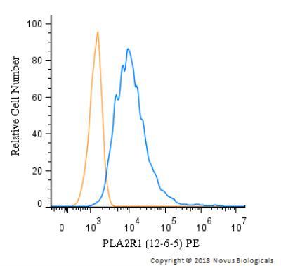
- Experimental details
- Flow Cytometry: PLA2R1 Antibody (12-6-5) [NBP2-50248] - A surface stain was performed on HeLa cells with PLA2R1 Antibody (12-6-5) NBP2-50248PE (blue) and a matched isotype control (orange). Cells were incubated in an antibody dilution of 2.5 ug/mL for 20 minutes at room temperature. Both antibodies were conjugated to phycoerythrin.