Explore
 Validate
 Learn
LearnPA5-81009
antibody from Invitrogen Antibodies
Targeting: SEMA6A
HT018, KIAA1368, SEMA, SEMA6A1, SEMAQ
 Western blot
 ELISA
ELISAAntibody data
- Antibody Data
- Antigen structure
- References [1]
- Comments [0]
- Validations
- Western blot [2]
- Immunocytochemistry [1]
Submit
Validation data
Reference
Comment
Report error
- Product number
- PA5-81009 - Provider product page

- Provider
- Invitrogen Antibodies
- Product name
- SEMA6A Polyclonal Antibody
- Antibody type
- Polyclonal
- Antigen
- Recombinant full-length protein
- Description
- This product is preservative free. It is recommended to add sodium azide to avoid contamination (final concentration 0.05%-0.1%). This antibody has specificity for Human SEMA6A.
- Reactivity
- Human
- Host
- Rabbit
- Isotype
- IgG
- Vial size
- 100 µL
- Concentration
- 1 mg/mL
- Storage
- Store at 4°C short term. For long term storage, store at -20°C, avoiding freeze/thaw cycles.
Submitted references Genome-Wide CRISPR Screen Identifies Semaphorin 6A and 6B as Receptors for Paeniclostridium sordellii Toxin TcsL.
Tian S, Liu Y, Wu H, Liu H, Zeng J, Choi MY, Chen H, Gerhard R, Dong M
Cell host & microbe 2020 May 13;27(5):782-792.e7
Cell host & microbe 2020 May 13;27(5):782-792.e7
No comments: Submit comment
Supportive validation
- Submitted by
- Invitrogen Antibodies (provider)
- Main image

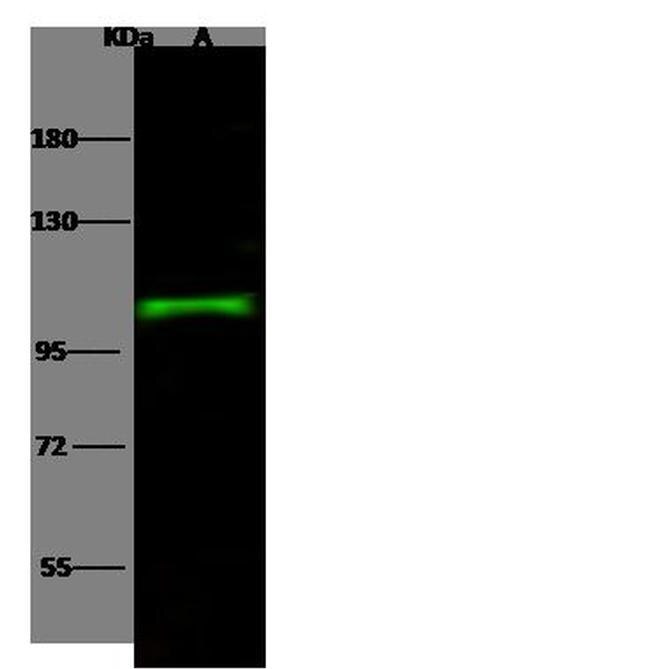
- Experimental details
- Western blot analysis of SEMA6A in Lane A: Raji Whole Cell Lysate (30 µg). Samples were probed using a SEMA6A Polyclonal Antibody (Product # PA5-81009) at a 1:500 dilution, followed by a Goat Anti-Rabbit IgG (H+L), Dylight 800 Secondary Antibody at a 1:10000 dilution. Western blot was performed under reducing conditions. Predicted band size:114 kDa. Observed band size:107 kDa.
- Submitted by
- Invitrogen Antibodies (provider)
- Main image
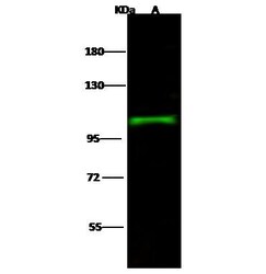
- Experimental details
- Western Blot using SEMA6A Polyclonal Antibody (Product # PA5-81009) at 1:500 dilution. Lane A: Raji Whole Cell Lysate. Lysates/proteins at 30 μg per lane. Secondary Goat Anti- RabbitIgG H&L (DyLight™ 800) at 1:10,000 dilution. Developed using the Odyssey technique. Performed under reducing conditions. Predicted band size: 114 kDa. Observed band size: 107 kDa.

Supportive validation
- Submitted by
- Invitrogen Antibodies (provider)
- Main image

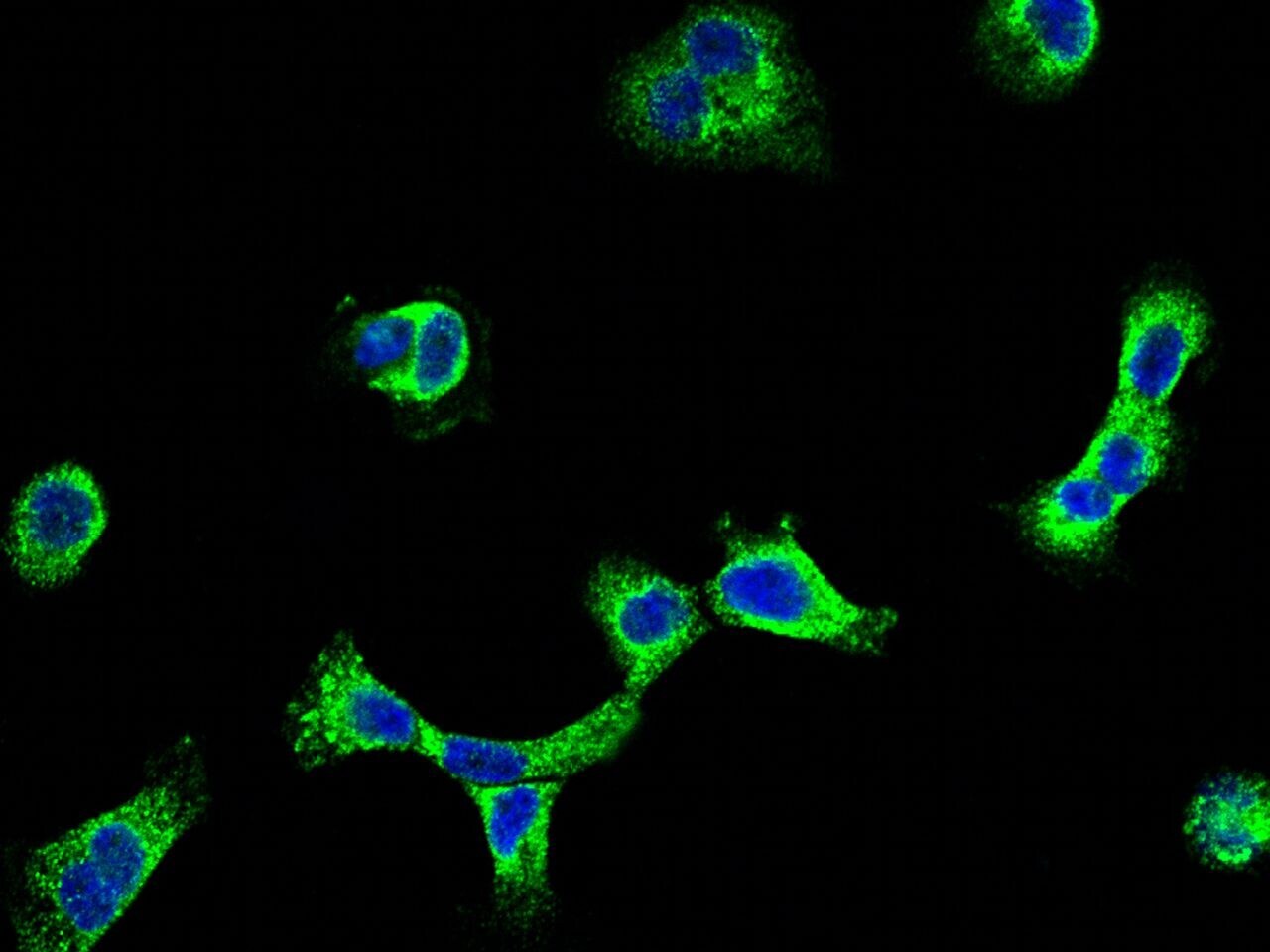
- Experimental details
- Immunofluorescence staining of SEMA6A in A431 cells. Cells were fixed with 4% PFA, permeabilzed with 0.3% Triton X-100 in PBS, blocked with 10% serum, and incubated with SEMA6A Polyclonal Antibody (Product # PA5-81009, 1:5,000) at 4°C overnight. Then cells were stained with the Alexa Fluor®488-conjugated Goat Anti-rabbit IgG secondary antibody (green) and counterstained with DAPI (blue). Positive staining was localized to cytoplasm.