 Explore
 Validate
 Learn
Learn Western blot
 Immunocytochemistry
Antibody data
- Antibody Data
- Antigen structure
- References [1]
- Comments [0]
- Validations
- Western blot [1]
- Immunocytochemistry [1]
- Immunohistochemistry [7]
Submit
Validation data
Reference
Comment
Report error
- Product number
- HPA001078 - Provider product page

- Provider
- Atlas Antibodies
- Proper citation
- Atlas Antibodies Cat#HPA001078, RRID:AB_10602432
- Product name
- Anti-APOL2
- Antibody type
- Polyclonal
- Reactivity
- Human
- Host
- Rabbit
- Conjugate
- Unconjugated
- Antigen sequence
LGVRVREEEAGTRVKENLPVWTVTGELQGKPLGNP
AAGTMNPESSIFIEDYLKYFQDQVSRENLLQLLTD
DEAWNGFVAAAELPRDEADELRKALNKLASHMVMK
DKNRHDKDQQHRQWFLKEFPRLKRELEDHIRKLRA
LAEEVEQVHR- Isotype
- IgG
- Vial size
- 100 µl
- Storage
- Store at +4°C for short term storage. Long time storage is recommended at -20°C.
Submitted references Apolipoprotein L2 contains a BH3-like domain but it does not behave as a BH3-only protein.
Galindo-Moreno J, Iurlaro R, El Mjiyad N, Díez-Pérez J, Gabaldón T, Muñoz-Pinedo C
Cell death & disease 2014 Jun 5;5(6):e1275
Cell death & disease 2014 Jun 5;5(6):e1275
No comments: Submit comment
Enhanced validation
- Submitted by
- Atlas Antibodies (provider)
- Enhanced method
- Genetic validation
- Main image
- Experimental details
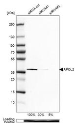
- Western blot analysis in U-138MG cells transfected with control siRNA, target specific siRNA probe #1 and #2, using Anti-APOL2 antibody. Remaining relative intensity is presented. Loading control: Anti-PPIB.

Supportive validation
- Submitted by
- Atlas Antibodies (provider)
- Main image
- Experimental details
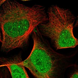
- Immunofluorescent staining of human cell line U-2 OS shows localization to nucleoplasm, nuclear bodies & cytosol.
- Sample type
- HUMAN

Enhanced validation
Supportive validation
- Submitted by
- Atlas Antibodies (provider)
- Enhanced method
- Orthogonal validation
- Main image
- Experimental details
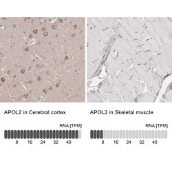
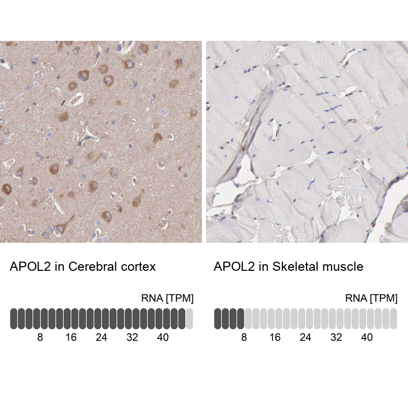
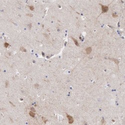
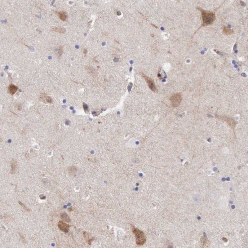
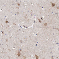
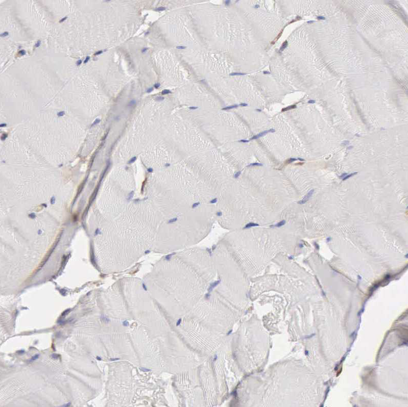
- Immunohistochemistry analysis in human cerebral cortex and skeletal muscle tissues using HPA001078 antibody. Corresponding APOL2 RNA-seq data are presented for the same tissues.
- Sample type
- HUMAN
Supportive validation
- Submitted by
- Atlas Antibodies (provider)
- Main image
- Experimental details
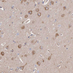
- Immunohistochemical staining of human hippocampus shows strong cytoplasmic and weak nuclear positivity in neuronal cells.
- Submitted by
- Atlas Antibodies (provider)
- Main image
- Experimental details
- Immunohistochemical staining of human hippocampus shows moderate cytoplasmic positivity in neuronal cells.
- Sample type
- HUMAN

- Submitted by
- Atlas Antibodies (provider)
- Main image

- Experimental details
- Immunohistochemical staining of human endometrium shows moderate cytoplasmic positivity in glandular cells.
- Sample type
- HUMAN

- Submitted by
- Atlas Antibodies (provider)
- Main image

- Experimental details
- Immunohistochemical staining of human liver shows moderate cytoplasmic positivity in hepatocytes.
- Sample type
- HUMAN

- Submitted by
- Atlas Antibodies (provider)
- Main image

- Experimental details
- Immunohistochemical staining of human skeletal muscle shows no positivity in myocytes as expected.
- Sample type
- HUMAN
- Submitted by
- Atlas Antibodies (provider)
- Main image
- Experimental details
- Immunohistochemical staining of human cerebral cortex shows moderate cytoplasmic positivity in neuronal cells.
- Sample type
- HUMAN
