Explore
 Validate
 Learn
Learn Western blot
Antibody data
- Antibody Data
- Antigen structure
- References [1]
- Comments [0]
- Validations
- Western blot [1]
- Other assay [2]
Submit
Validation data
Reference
Comment
Report error
- Product number
- PA5-28809 - Provider product page

- Provider
- Invitrogen Antibodies
- Product name
- HIPK3 Polyclonal Antibody
- Antibody type
- Polyclonal
- Antigen
- Synthetic peptide
- Description
- Recommended positive controls: A431, H1299, HeLa, HepG2.
- Concentration
- 0.3 mg/mL
Submitted references Hsa_circ_0025202 suppresses cell tumorigenesis and tamoxifen resistance via miR-197-3p/HIPK3 axis in breast cancer.
Li H, Li Q, He S
World journal of surgical oncology 2021 Feb 3;19(1):39
World journal of surgical oncology 2021 Feb 3;19(1):39
No comments: Submit comment
Supportive validation
- Submitted by
- Invitrogen Antibodies (provider)
- Main image
- Experimental details
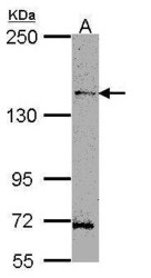
- Western Blot using HIPK3 Polyclonal Antibody (Product # PA5-28809). Sample (30 µg of whole cell lysate). Lane A: H1299 . 5% SDS PAGE. HIPK3 Polyclonal Antibody (Product # PA5-28809) diluted at 1:1,000.

Supportive validation
- Submitted by
- Invitrogen Antibodies (provider)
- Main image

- Experimental details
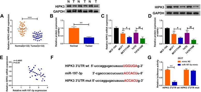
- Fig. 6 HIPK3 is a target of miR-197-3p. a , b qRT-PCR and western blot analysis of HIPK3 expression in BC tissues and matched non-tumor tissues. c , d qRT-PCR and western blot analysis of HIPK3 expression in normal MCF-10A cells, BC cell lines (T47D and MCF7), and TAM-resistant BC cell lines (T47D/TAM and MCF7/TAM). e The correlation between HIPK3 level and miR-197-3p expression in BC tissues using the Pearson correlation analysis. f The potential binding sites of HIPK3 and miR-197-3p. g Luciferase activities detection in 293 T cells co-transfected with the reporter plasmids and indicated miRNAs using the dual-luciferase reporter assay. ** P < 0.01, *** P < 0.001, # P < 0.05, ## P < 0.01
- Submitted by
- Invitrogen Antibodies (provider)
- Main image
- Experimental details
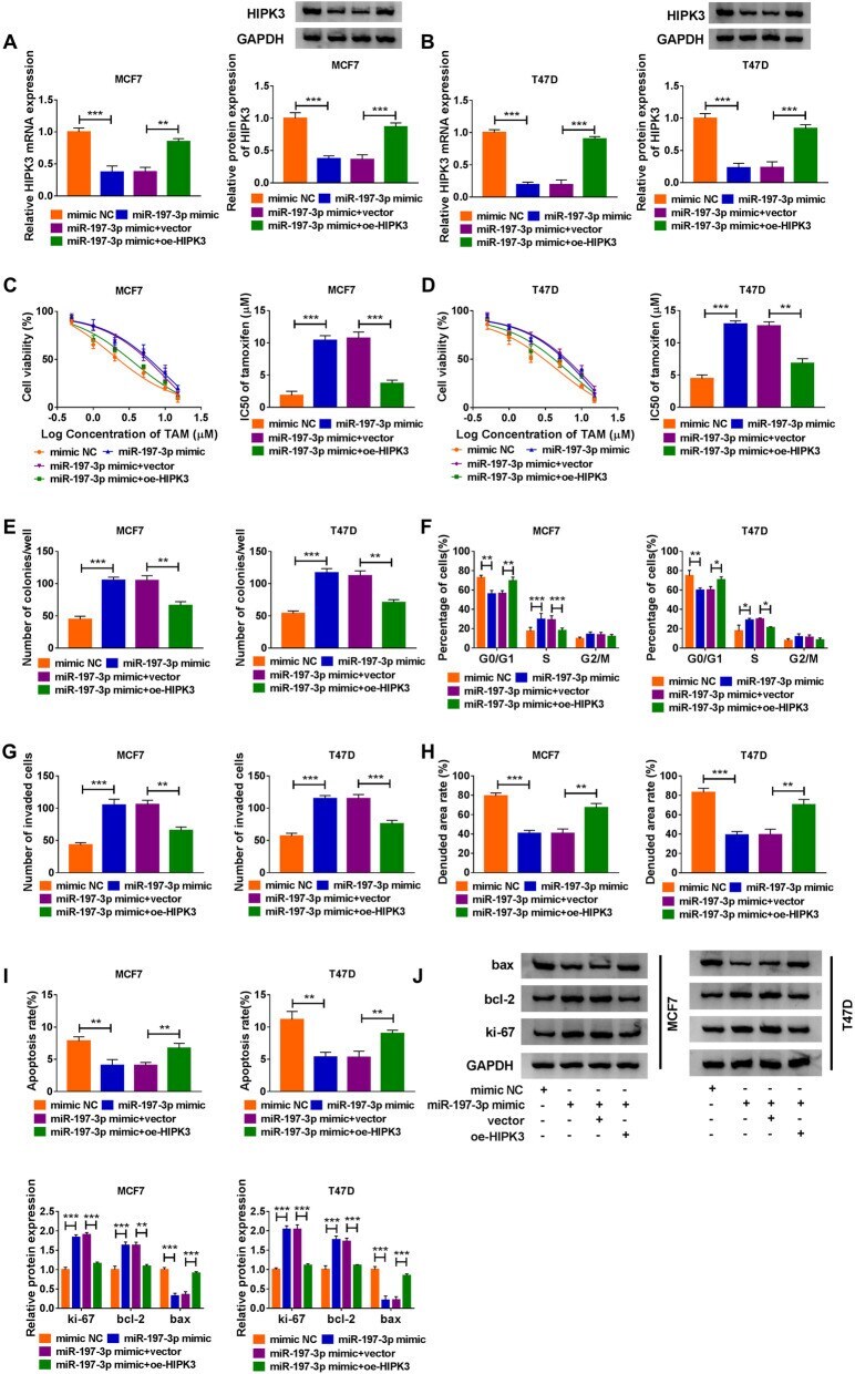
- Fig. 7 MiR-197-3p promotes cell TAM resistance and carcinogenesis in BC via HIPK3. a , b HIPK3 level by qRT-PCR and western blot; c , d the IC 50 value of TAM by CCK-8 assay; e cell proliferation by colony formation assay; f cell cycle by flow cytometry assay; g cell invasion by transwell assay; h cell migration by wound healing assay; i cell apoptosis by flow cytometry; j protein levels of bax, ki-67, and bcl-2 by western blot, in T47D and MCF7 cells transfected with mimic NC, miR-197-3p mimic, miR-197-3p mimic + vector, or miR-197-3p mimic + oe-HIPK3. * P < 0.05, ** P < 0.01, *** P < 0.001