Explore
 Validate
 Learn
Learn Western blot
Antibody data
- Antibody Data
- Antigen structure
- References [3]
- Comments [0]
- Validations
- Western blot [3]
- Immunocytochemistry [1]
- Immunohistochemistry [5]
Submit
Validation data
Reference
Comment
Report error
- Product number
- HPA003624 - Provider product page

- Provider
- Atlas Antibodies
- Proper citation
- Atlas Antibodies Cat#HPA003624, RRID:AB_10602123
- Product name
- Anti-RBM3
- Antibody type
- Polyclonal
- Reactivity
- Human, Mouse, Rat
- Host
- Rabbit
- Conjugate
- Unconjugated
- Antigen sequence
DEQALEDHFSSFGPISEVVVVKDRETQRSRGFGFI
TFTNPEHASVAMRAMNGESLDGRQIRVDHAGKSAR
GTRGGGFGAHGRGRSYSRGGGDQGYGSGRYYDSRP
GGYGYGYGRSRDYNGRNQGGYDRYSGGNY- Isotype
- IgG
- Vial size
- 100 µl
- Storage
- Store at +4°C for short term storage. Long time storage is recommended at -20°C.
Submitted references Generation of monospecific antibodies based on affinity capture of polyclonal antibodies
High nuclear RBM3 expression is associated with an improved prognosis in colorectal cancer
Nuclear expression of the RNA-binding protein RBM3 is associated with an improved clinical outcome in breast cancer
Hjelm B, Forsström B, Igel U, Johannesson H, Stadler C, Lundberg E, Ponten F, Sjöberg A, Rockberg J, Schwenk J, Nilsson P, Johansson C, Uhlén M
Protein Science 2011 November;20(11):1824-1835
Protein Science 2011 November;20(11):1824-1835
High nuclear RBM3 expression is associated with an improved prognosis in colorectal cancer
Hjelm B, Brennan D, Zendehrokh N, Eberhard J, Nodin B, Gaber A, Pontén F, Johannesson H, Smaragdi K, Frantz C, Hober S, Johnson L, Påhlman S, Jirström K, Uhlen M
PROTEOMICS - Clinical Applications 2011 December;5(11-12):624-635
PROTEOMICS - Clinical Applications 2011 December;5(11-12):624-635
Nuclear expression of the RNA-binding protein RBM3 is associated with an improved clinical outcome in breast cancer
Jögi A, Brennan D, Rydén L, Magnusson K, Fernö M, Stål O, Borgquist S, Uhlen M, Landberg G, Påhlman S, Pontén F, Jirström K
Modern Pathology 2009 September;22(12):1564-1574
Modern Pathology 2009 September;22(12):1564-1574
No comments: Submit comment
Supportive validation
- Submitted by
- Atlas Antibodies (provider)
- Enhanced method
- Genetic validation
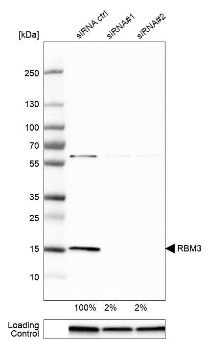
- Main image

- Experimental details
- Western blot analysis in U-251MG cells transfected with control siRNA, target specific siRNA probe #1 and #2, using Anti-RBM3 antibody. Remaining relative intensity is presented. Loading control: Anti-GAPDH.
- Submitted by
- Atlas Antibodies (provider)
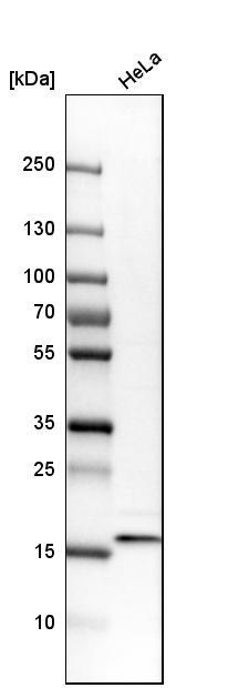
- Main image
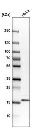
- Experimental details
- Western blot analysis in human cell line HeLa.
- Sample type
- HUMAN
- Submitted by
- Atlas Antibodies (provider)
- Main image
- Experimental details
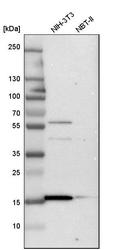
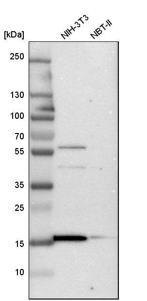
- Western blot analysis in mouse cell line NIH-3T3 and rat cell line NBT-II.
Supportive validation
- Submitted by
- Atlas Antibodies (provider)
- Main image

- Experimental details
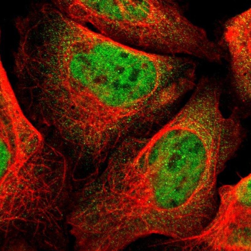
- Immunofluorescent staining of human cell line U-2 OS shows localization to nucleoplasm.
- Sample type
- HUMAN
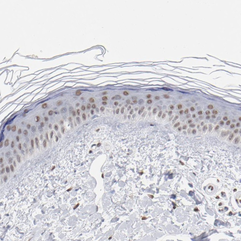
Supportive validation
- Submitted by
- Atlas Antibodies (provider)
- Main image
- Experimental details
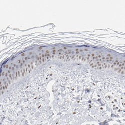
- Immunohistochemical staining of human skin shows moderate nuclear positivity in epidermal cells.
- Sample type
- HUMAN
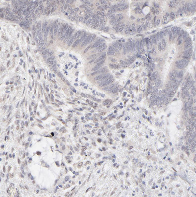
- Submitted by
- Atlas Antibodies (provider)
- Main image
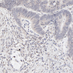
- Experimental details
- Immunohistochemical staining of human colorectal cancer shows weak nuclear positivity in tumor cells.
- Sample type
- HUMAN
- Submitted by
- Atlas Antibodies (provider)
- Main image
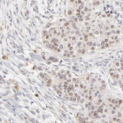
- Experimental details
- Immunohistochemical staining of human breast cancer shows moderate nuclear positivity in tumor cells.
- Sample type
- HUMAN

- Submitted by
- Atlas Antibodies (provider)
- Main image
- Experimental details
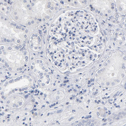
- Immunohistochemical staining of human kidney shows no nuclear positivity in cells in glomeruli as expected.
- Sample type
- HUMAN

- Submitted by
- Atlas Antibodies (provider)
- Main image
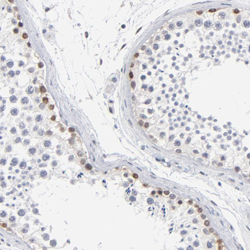
- Experimental details
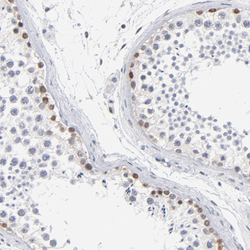
- Immunohistochemical staining of human testis shows moderate nuclear positivity in cells in seminiferous ducts.
- Sample type
- HUMAN