 Explore
 Validate
 Learn
Learn Western blot
 Immunohistochemistry
Antibody data
- Antibody Data
- Antigen structure
- References [0]
- Comments [0]
- Validations
- Western blot [3]
- Immunohistochemistry [7]
Submit
Validation data
Reference
Comment
Report error
- Product number
- AMAb91245 - Provider product page

- Provider
- Atlas Antibodies
- Proper citation
- Atlas Antibodies Cat#AMAb91245, RRID:AB_2665861
- Product name
- Anti-PPIB
- Antibody type
- Monoclonal
- Antigen
- Synthetic peptide
- Reactivity
- Human, Mouse, Rat
- Host
- Mouse
- Conjugate
- Unconjugated
- Antigen sequence
TKTDSRDKPLKDVIIADCGKIEVEKPFAIAKE- Isotype
- IgG
- Antibody clone number
- CL3901
- Vial size
- 100 µl
- Storage
- Store at +4°C for short term storage. Long time storage is recommended at -20°C.
No comments: Submit comment
Enhanced validation
- Submitted by
- Atlas Antibodies (provider)
- Enhanced method
- Genetic validation
- Main image
- Experimental details
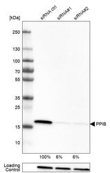
- Western blot analysis in U-251MG cells transfected with control siRNA, target specific siRNA probe #1 and #2, using Anti-PPIB antibody. Remaining relative intensity is presented. Loading control: Anti-GAPDH.

- Submitted by
- Atlas Antibodies (provider)
- Main image

- Experimental details
- Western blot analysis in human cell line HeLa, human cell line HEK 293, human cell line A-431, human cell line HepG2, mouse cell line NIH-3T3 and rat cell line NBT-II.

- Submitted by
- Atlas Antibodies (provider)
- Main image
- Experimental details
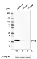
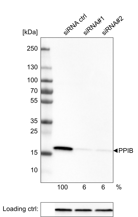
- Western blot analysis of extracts from U-251 cells, transfected with: control siRNA, target specific siRNA probe #1, target specific siRNA probe #2, using Anti-PPIB monoclonal antibody. Downregulation of antibody signal confirms target specificity. Remaining % intensity, relative control lane, is indicated. Anti-GAPDH monoclonal antibody was used as loading control.
Supportive validation
- Submitted by
- Atlas Antibodies (provider)
- Main image
- Experimental details
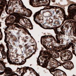
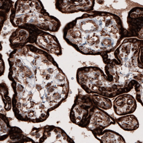
- Immunohistochemical staining of human placenta shows strong cytoplasmic immunoreactivity in trophoblasts.
- Submitted by
- Atlas Antibodies (provider)
- Main image
- Experimental details
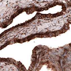
- Immunohistochemical staining of human prostate shows cytoplasmic positivity in glandular cells.

- Submitted by
- Atlas Antibodies (provider)
- Main image
- Experimental details
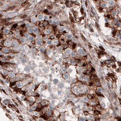
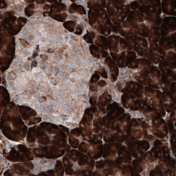
- Immunohistochemical staining of human testis shows strong cytoplasmic immunoreactivity in a subset of cells in seminiferous tubules.

- Submitted by
- Atlas Antibodies (provider)
- Main image
- Experimental details
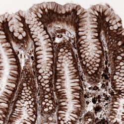
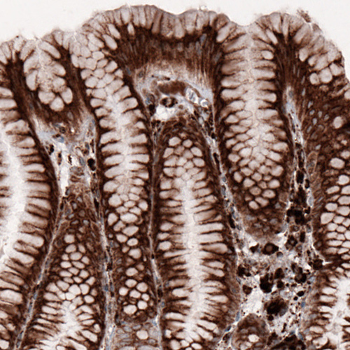
- Immunohistochemical staining of human stomach shows cytoplasmic positivity in glandular and lymphoid cells.
- Submitted by
- Atlas Antibodies (provider)
- Main image
- Experimental details
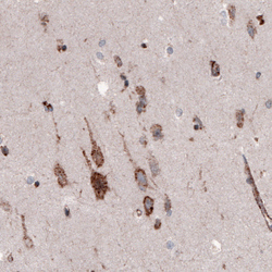
- Immunohistochemical staining of human cerebral cortex shows cytoplasmic immunoreactivity in neurons.

- Submitted by
- Atlas Antibodies (provider)
- Main image
- Experimental details
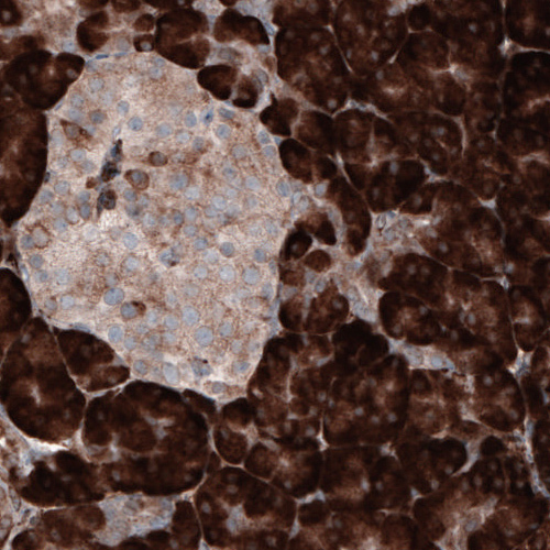
- Immunohistochemical staining of human pancreas shows strong cytoplasmic positivity in the exocrine cells and weak immunoreactivity in the endocrine cells.
- Submitted by
- Atlas Antibodies (provider)
- Main image
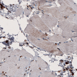
- Experimental details
- Immunohistochemical staining of human skeletal muscle shows very weak cytoplasmic positivity in muscle fibers.
