Explore
 Validate
 Learn
Learn Western blot
 Immunocytochemistry
Antibody data
- Antibody Data
- Antigen structure
- References [2]
- Comments [0]
- Validations
- Western blot [1]
- Immunocytochemistry [1]
- Immunohistochemistry [9]
Submit
Validation data
Reference
Comment
Report error
- Product number
- HPA012720 - Provider product page

- Provider
- Atlas Antibodies
- Proper citation
- Atlas Antibodies Cat#HPA012720, RRID:AB_1847399
- Product name
- Anti-PPIB
- Antibody type
- Polyclonal
- Reactivity
- Human, Mouse, Rat
- Host
- Rabbit
- Conjugate
- Unconjugated
- Antigen sequence
LKHYGPGWVSMANAGKDTNGSQFFITTVKTAWLDG
KHVVFGKVLEGMEVVRKVESTKTDSRDKPLKDVII
ADCGKIEVEKPFAIAKE- Isotype
- IgG
- Vial size
- 100 µl
- Storage
- Store at +4°C for short term storage. Long time storage is recommended at -20°C.
Submitted references Galectin-4, a novel predictor for lymph node metastasis in lung adenocarcinoma.
PPIB mutations cause severe osteogenesis imperfecta.
Hayashi T, Saito T, Fujimura T, Hara K, Takamochi K, Mitani K, Mineki R, Kazuno S, Oh S, Ueno T, Suzuki K, Yao T
PloS one 2013;8(12):e81883
PloS one 2013;8(12):e81883
PPIB mutations cause severe osteogenesis imperfecta.
van Dijk FS, Nesbitt IM, Zwikstra EH, Nikkels PG, Piersma SR, Fratantoni SA, Jimenez CR, Huizer M, Morsman AC, Cobben JM, van Roij MH, Elting MW, Verbeke JI, Wijnaendts LC, Shaw NJ, Högler W, McKeown C, Sistermans EA, Dalton A, Meijers-Heijboer H, Pals G
American journal of human genetics 2009 Oct;85(4):521-7
American journal of human genetics 2009 Oct;85(4):521-7
No comments: Submit comment
Enhanced validation
- Submitted by
- Atlas Antibodies (provider)
- Enhanced method
- Genetic validation
- Main image

- Experimental details
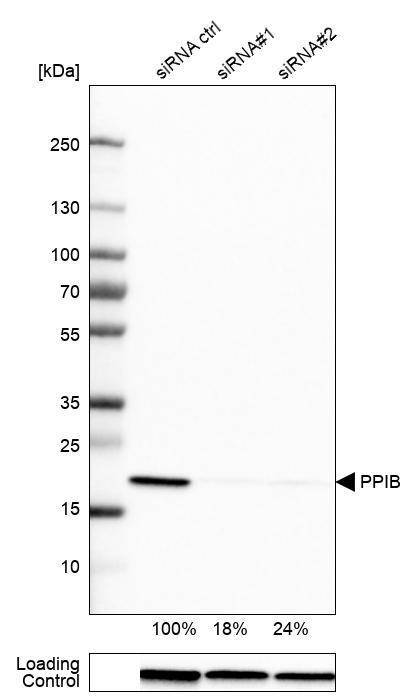
- Western blot analysis in U-251MG cells transfected with control siRNA, target specific siRNA probe #1 and #2, using Anti-PPIB antibody. Remaining relative intensity is presented. Loading control: Anti-GAPDH.
Supportive validation
- Submitted by
- Atlas Antibodies (provider)
- Main image
- Experimental details
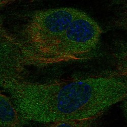
- Immunofluorescent staining of human cell line HUVEC TERT2 shows localization to endoplasmic reticulum.
- Sample type
- HUMAN

Enhanced validation
Supportive validation
- Submitted by
- Atlas Antibodies (provider)
- Enhanced method
- Orthogonal validation
- Main image
- Experimental details
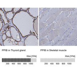
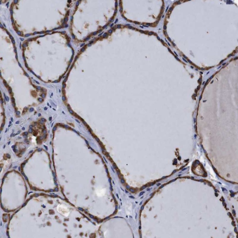
- Immunohistochemistry analysis in human thyroid gland and skeletal muscle tissues using HPA012720 antibody. Corresponding PPIB RNA-seq data are presented for the same tissues.
- Sample type
- HUMAN

Supportive validation
- Submitted by
- Atlas Antibodies (provider)
- Main image
- Experimental details
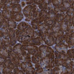
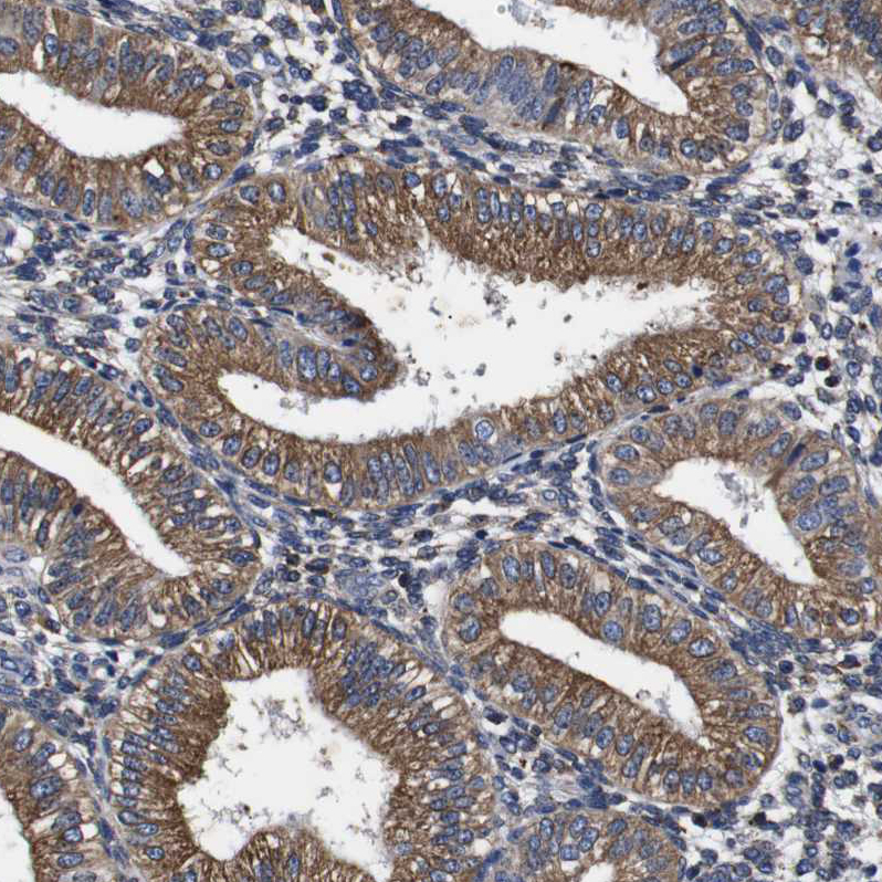
- Immunohistochemical staining of human pancreas shows strong cytoplasmic positivity in exocrine glandular cells.
- Submitted by
- Atlas Antibodies (provider)
- Main image

- Experimental details
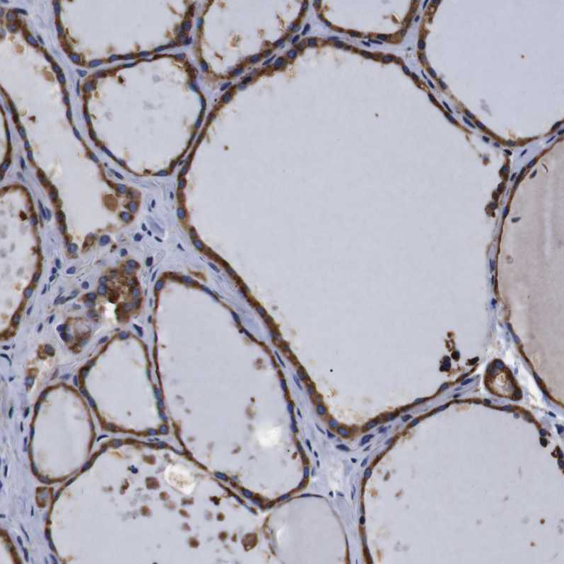
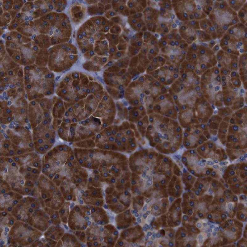
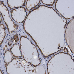
- Immunohistochemical staining of human thyroid gland shows high expression.
- Sample type
- HUMAN
- Submitted by
- Atlas Antibodies (provider)
- Main image
- Experimental details
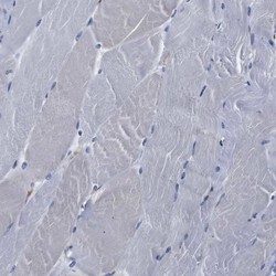
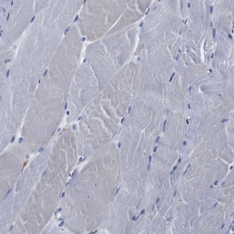
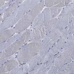
- Immunohistochemical staining of human skeletal muscle shows low expression as expected.
- Sample type
- HUMAN
- Submitted by
- Atlas Antibodies (provider)
- Main image
- Experimental details
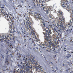
- Immunohistochemical staining of human prostate shows weak granular cytoplasmic positivity in glandular cells.
- Sample type
- HUMAN

- Submitted by
- Atlas Antibodies (provider)
- Main image
- Experimental details
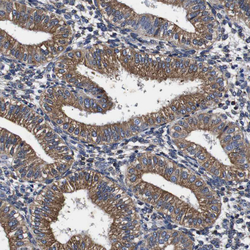
- Immunohistochemical staining of human endometrium shows moderate granular cytoplasmic positivity in glandular cells.
- Sample type
- HUMAN
- Submitted by
- Atlas Antibodies (provider)
- Main image
- Experimental details
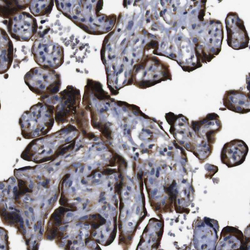
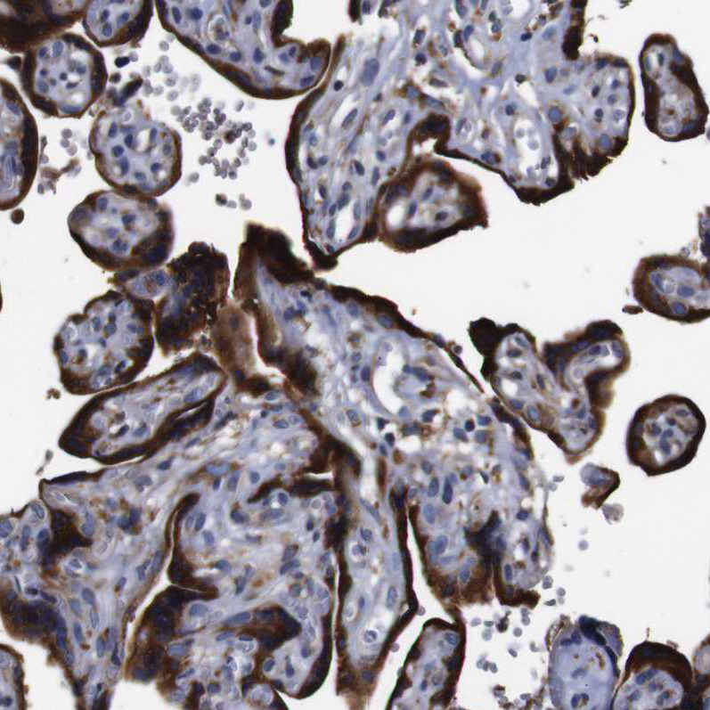
- Immunohistochemical staining of human placenta shows strong granular cytoplasmic positivity in trophoblastic cells.
- Sample type
- HUMAN
- Submitted by
- Atlas Antibodies (provider)
- Main image
- Experimental details
- Immunohistochemical staining of human skeletal muscle shows no positivity in myocytes as expected.
- Sample type
- HUMAN

- Submitted by
- Atlas Antibodies (provider)
- Main image
- Experimental details
- Immunohistochemical staining of human thyroid gland shows strong granular cytoplasmic positivity in glandular cells.
- Sample type
- HUMAN