 Explore
 Validate
 Learn
Learn Western blot
Antibody data
- Antibody Data
- Antigen structure
- References [1]
- Comments [0]
- Validations
- Western blot [4]
- Immunohistochemistry [9]
Submit
Validation data
Reference
Comment
Report error
- Product number
- TA500408 - Provider product page

- Provider
- OriGene
- Proper citation
- OriGene Cat#TA500408, RRID:AB_2196462
- Product name
- Anti-STK3 mouse monoclonal antibody, clone OTI4G10 (formerly 4G10)
- Antibody type
- Monoclonal
- Description
- Anti-STK3 mouse monoclonal antibody, clone OTI4G10 (formerly 4G10)
- Reactivity
- Canine
- Host
- Mouse
- Conjugate
- Unconjugated
- Epitope
- STK3
- Isotype
- IgG
- Antibody clone number
- OTI4G10
- Vial size
- 100 µl
- Concentration
- 1 mg/ml
Submitted references Poly(rC)-Binding Protein 2 Regulates Hippo Signaling To Control Growth in Breast Epithelial Cells.
Li F, Bullough KZ, Vashisht AA, Wohlschlegel JA, Philpott CC
Molecular and cellular biology 2016 Aug 15;36(16):2121-31
Molecular and cellular biology 2016 Aug 15;36(16):2121-31
No comments: Submit comment
Supportive validation
- Submitted by
- OriGene (provider)
- Main image

- Experimental details
- HEK293T cells were transfected with the pCMV6-ENTRY control (Left lane) or pCMV6-ENTRY STK3 (RC203897, Right lane) cDNA for 48 hrs and lysed. Equivalent amounts of cell lysates (5 ug per lane) were separated by SDS-PAGE and immunoblotted with anti-STK3.
- Validation comment
- WB

- Submitted by
- OriGene (provider)
- Main image
- Experimental details
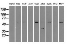
- Western blot analysis of extracts (35ug) from 9 different cell lines by using anti-STK3 monoclonal antibody.
- Validation comment
- WB

- Submitted by
- OriGene (provider)
- Main image
- Experimental details
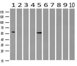
- Western blot analysis of extracts (10ug) from 10 Human tissue by using anti-STK3 monoclonal antibody at 1:200 (1: Testis; 2: Omentum; 3: Uterus; 4: Breast; 5: Brain; 6: Liver; 7: Ovary; 8: Thyroid gland; 9: colon;10: spleen).
- Validation comment
- WB

- Submitted by
- OriGene (provider)
- Main image
- Experimental details
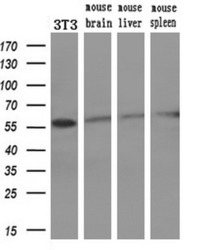
- Western blot analysis of extracts (10ug) from a mouse cell line and 3 different mouse tissues by using anti-STK3 monoclonal antibody. (1:200)
- Validation comment
- WB

Supportive validation
- Submitted by
- OriGene (provider)
- Main image

- Experimental details
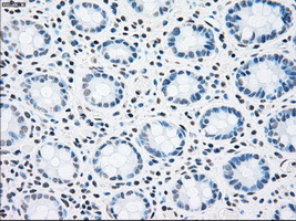
- Immunohistochemical staining of paraffin-embedded colon tissue within the normal limits using anti-STK3mouse monoclonal antibody. (Heat-induced epitope retrieval by 10mM citric buffer, pH6.0, 100C for 10min, TA500408, Dilution 1:50)
- Validation comment
- IHC
- Submitted by
- OriGene (provider)
- Main image

- Experimental details
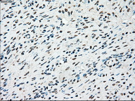
- Immunohistochemical staining of paraffin-embedded Adenocarcinoma of colon tissue using anti-STK3mouse monoclonal antibody. (Heat-induced epitope retrieval by 10mM citric buffer, pH6.0, 100C for 10min, TA500408, Dilution 1:50)
- Validation comment
- IHC

- Submitted by
- OriGene (provider)
- Main image

- Experimental details
- Immunohistochemical staining of paraffin-embedded Kidney tissue within the normal limits using anti-STK3mouse monoclonal antibody. (Heat-induced epitope retrieval by 10mM citric buffer, pH6.0, 100C for 10min, TA500408, Dilution 1:50)
- Validation comment
- IHC

- Submitted by
- OriGene (provider)
- Main image

- Experimental details
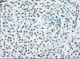
- Immunohistochemical staining of paraffin-embedded Carcinoma of kidney tissue using anti-STK3mouse monoclonal antibody. (Heat-induced epitope retrieval by 10mM citric buffer, pH6.0, 100C for 10min, TA500408, Dilution 1:50)
- Validation comment
- IHC

- Submitted by
- OriGene (provider)
- Main image

- Experimental details
- Immunohistochemical staining of paraffin-embedded liver tissue within the normal limits using anti-STK3mouse monoclonal antibody. (Heat-induced epitope retrieval by 10mM citric buffer, pH6.0, 100C for 10min, TA500408, Dilution 1:50)
- Validation comment
- IHC

- Submitted by
- OriGene (provider)
- Main image

- Experimental details
- Immunohistochemical staining of paraffin-embedded lung tissue within the normal limits using anti-STK3mouse monoclonal antibody. (Heat-induced epitope retrieval by 10mM citric buffer, pH6.0, 100C for 10min, TA500408, Dilution 1:50)
- Validation comment
- IHC

- Submitted by
- OriGene (provider)
- Main image

- Experimental details
- Immunohistochemical staining of paraffin-embedded Ovary tissue within the normal limits using anti-STK3mouse monoclonal antibody. (Heat-induced epitope retrieval by 10mM citric buffer, pH6.0, 100C for 10min, TA500408, Dilution 1:50)
- Validation comment
- IHC
- Submitted by
- OriGene (provider)
- Main image

- Experimental details
- Immunohistochemical staining of paraffin-embedded pancreas tissue within the normal limits using anti-STK3mouse monoclonal antibody. (Heat-induced epitope retrieval by 10mM citric buffer, pH6.0, 100C for 10min, TA500408, Dilution 1:50)
- Validation comment
- IHC
- Submitted by
- OriGene (provider)
- Main image

- Experimental details
- Immunohistochemical staining of paraffin-embedded endometrium tissue within the normal limits using anti-STK3mouse monoclonal antibody. (Heat-induced epitope retrieval by 10mM citric buffer, pH6.0, 100C for 10min, TA500408, Dilution 1:50)
- Validation comment
- IHC
