Explore
 Validate
 Learn
Learn Western blot
Antibody data
- Antibody Data
- Antigen structure
- References [1]
- Comments [0]
- Validations
- Western blot [7]
- Immunocytochemistry [4]
Submit
Validation data
Reference
Comment
Report error
- Product number
- PA5-21142 - Provider product page

- Provider
- Invitrogen Antibodies
- Product name
- SPTLC2 Polyclonal Antibody
- Antibody type
- Polyclonal
- Antigen
- Synthetic peptide
- Description
- A suggested positive control is NIH-3T3 cell lysate.
- Concentration
- 1 mg/mL
Submitted references Increased de novo ceramide synthesis and accumulation in failing myocardium.
Ji R, Akashi H, Drosatos K, Liao X, Jiang H, Kennel PJ, Brunjes DL, Castillero E, Zhang X, Deng LY, Homma S, George IJ, Takayama H, Naka Y, Goldberg IJ, Schulze PC
JCI insight 2017 May 4;2(9)
JCI insight 2017 May 4;2(9)
No comments: Submit comment
Supportive validation
- Submitted by
- Invitrogen Antibodies (provider)
- Main image
- Experimental details
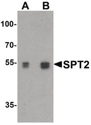
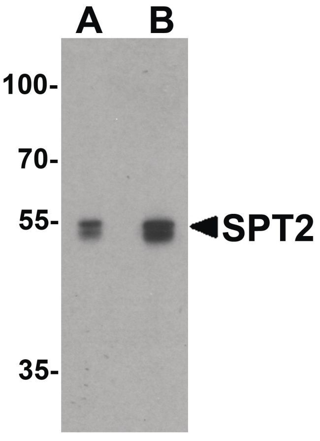
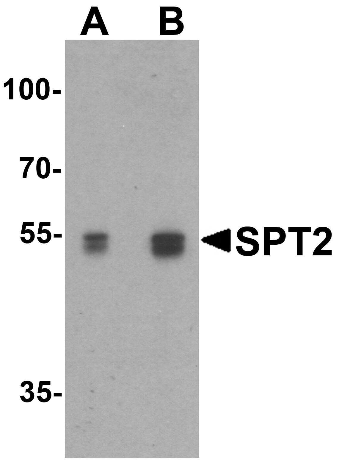
- Western blot analysis of 3T3 cell lysate using a SPT2 polyclonal antibody (Product # PA5-21142) at (A) 0.25 and (B) 0.5 µg/mL.
- Submitted by
- Invitrogen Antibodies (provider)
- Main image
- Experimental details
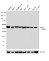
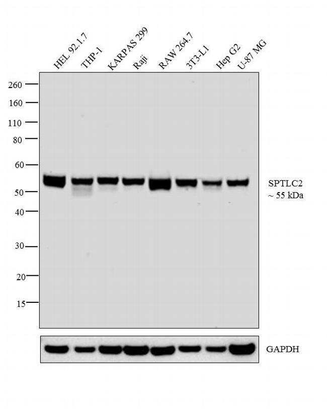
- Western blot analysis was performed on whole cell extracts (30 µg lysate) of HEL 92.1.7 (Lane 1), THP-1 (Lane 2), KARPAS 299 (Lane 3), Raji (Lane 4), RAW 264.7 (Lane 5), 3T3-L1 (Lane 6), Hep G2 (Lane 7) and U-87 MG (Lane 8). The blot was probed with Anti-SPTLC2 Polyclonal Antibody (Product # PA5-21142, 1 µg/ml) and detected by chemiluminescence using Goat anti-Rabbit IgG (H+L) Superclonal™ Secondary Antibody, HRP conjugate (Product # A27036, 0.25 µg/ml, 1:4000 dilution). A 55 kDa band corresponding to SPTLC2 was observed across the cell lines tested.
- Submitted by
- Invitrogen Antibodies (provider)
- Main image
- Experimental details
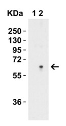
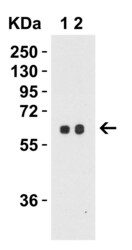
- Western Blot Validation in Human Brain Tissue Lysate. Loading: 15 µg of lysates per lane. Antibodies: SPTLC2 Polyclonal Antibody (Product # PA5-21142) (Lane 1: 0.5 µg/mL and Lane 2: 1 µg/mL), 1h incubation at RT in 0.05 NFDM/TBST. Secondary: Goat anti-rabbit IgG HRP conjugate at 1:10,000 dilution.

- Submitted by
- Invitrogen Antibodies (provider)
- Main image
- Experimental details
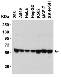
- Western Blot Validation in Human Cell Lines. Loading: 15 µg of lysates per lane. Antibodies: SPTLC2 Polyclonal Antibody (Product # PA5-21142) (1 µg/mL), 1h incubation at RT in 0.05 NFDM/TBST. Secondary: Goat anti-rabbit IgG HRP conjugate at 1:10,000 dilution.

- Submitted by
- Invitrogen Antibodies (provider)
- Main image

- Experimental details
- Western Blot Validation in Mouse 3T3 Cell Lysate in (A) the absence and (B) the presence of blocking peptide. Loading: 15 µg of lysates per lane. Antibodies: SPTLC2 Polyclonal Antibody (Product # PA5-21142) (0.5 µg/mL), 1h incubation at RT in 0.05 NFDM/TBST. Secondary: Goat anti-rabbit IgG HRP conjugate at 1:10,000 dilution.
- Submitted by
- Invitrogen Antibodies (provider)
- Main image
- Experimental details
- Western Blot Validation in Mouse 3T3/NIH Cell Lysate. Loading: 15 µg of lysates per lane. Antibodies: SPTLC2 Polyclonal Antibody (Product # PA5-21142) (Lane 1: 0.5 µg/mL and Lane 2: 1 µg/mL), 1h incubation at RT in 0.05 NFDM/TBST. Secondary: Goat anti-rabbit IgG HRP conjugate at 1:10,000 dilution.

- Submitted by
- Invitrogen Antibodies (provider)
- Main image
- Experimental details
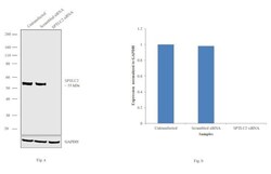
- Knockdown of SPTLC2 was achieved by transfecting U-87 MG cells with SPTLC2 specific siRNAs (Silencer® select Product # s225153). Western blot analysis (Fig. a) was performed using membrane enriched extracts from the SPTLC2 knockdown cells (lane 3), non-specific scrambled siRNA transfected cells (lane 2) and untransfected cells (lane 1). The blot was probed with SPTLC2 Polyclonal Antibody (Product # PA5-21142, 1:1000 dilution) and Goat anti-Rabbit IgG (H+L) Superclonal™ Secondary Antibody, HRP conjugate (Product # A27036, 0.25µg/ml, 1:4000 dilution). Densitometric analysis of this western blot is shown in histogram (Fig. b). Decrease in signal upon siRNA mediated knock down confirms that antibody is specific to SPTLC2.

Supportive validation
- Submitted by
- Invitrogen Antibodies (provider)
- Main image

- Experimental details
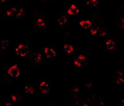
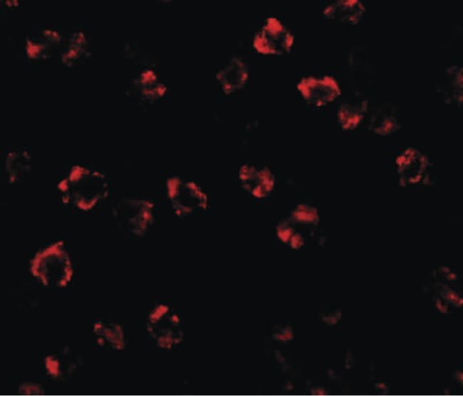
- Immunofluorescent analysis of 3T3 cells using a SPT2 polyclonal antibody (Product # PA5-21142) at a 20 µg/mL dilution.
- Submitted by
- Invitrogen Antibodies (provider)
- Main image

- Experimental details
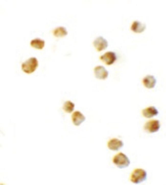
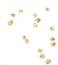
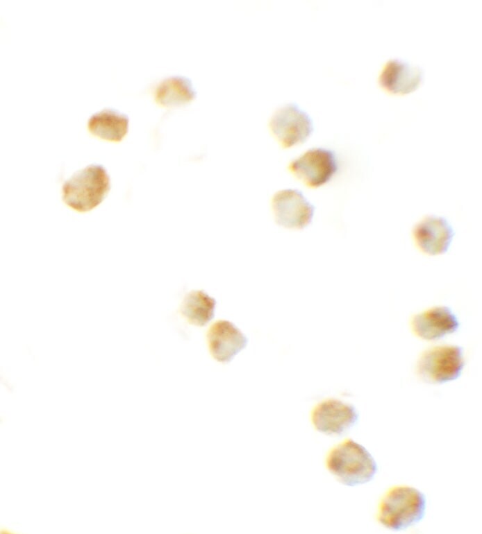
- Immunocytochemistry staining of 3T3 cells using a SPT2 polyclonal antibody (Product # PA5-21142) at a 10 µg/mL dilution.
- Submitted by
- Invitrogen Antibodies (provider)
- Main image
- Experimental details
- Immunocytochemistry of 3T3 cells using SPTLC2 Polyclonal Antibody (Product # PA5-21142) at 10 µg/mL. Cells were fixed with formaldehyde and blocked with 0.1 serum for 1 h at RT; antigen retrieval was by heat mediation with a citrate buffer (pH6). Samples were incubated with primary antibody overnight at 4°C. A goat anti-rabbit IgG H&L (HRP) at 1:250 was used as secondary. Counter stained with Hematoxylin.
- Submitted by
- Invitrogen Antibodies (provider)
- Main image

- Experimental details
- Immunofluorescent analysis of 4% paraformaldehyde-fixed 3T3 Cells labeling SPT2 with SPTLC2 Polyclonal Antibody (Product # PA5-21142) at 20 µg/mL, followed by goat anti-rabbit IgG secondary antibody at 1:500 dilution (red).