Antibody data
- Antibody Data
- Antigen structure
- References [1]
- Comments [0]
- Validations
- Western blot [2]
- Immunocytochemistry [2]
- Immunohistochemistry [1]
- Other assay [2]
Submit
Validation data
Reference
Comment
Report error
- Product number
- PA5-31356 - Provider product page

- Provider
- Invitrogen Antibodies
- Product name
- Septin-8 Polyclonal Antibody
- Antibody type
- Polyclonal
- Antigen
- Recombinant protein fragment
- Description
- Recommended positive controls: HCT116, mouse brain, rat brain. Predicted reactivity: Mouse (98%), Rat (98%), Xenopus laevis (87%), Rabbit (96%), Chicken (90%), Bovine (99%). Store product as a concentrated solution. Centrifuge briefly prior to opening the vial.
- Reactivity
- Human, Mouse, Rat
- Host
- Rabbit
- Isotype
- IgG
- Vial size
- 100 µL
- Concentration
- 0.83 mg/mL
- Storage
- Store at 4°C short term. For long term storage, store at -20°C, avoiding freeze/thaw cycles.
Submitted references Sept8/SEPTIN8 involvement in cellular structure and kidney damage is identified by genetic mapping and a novel human tubule hypoxic model.
Keele GR, Prokop JW, He H, Holl K, Littrell J, Deal AW, Kim Y, Kyle PB, Attipoe E, Johnson AC, Uhl KL, Sirpilla OL, Jahanbakhsh S, Robinson M, Levy S, Valdar W, Garrett MR, Solberg Woods LC
Scientific reports 2021 Jan 22;11(1):2071
Scientific reports 2021 Jan 22;11(1):2071
No comments: Submit comment
Supportive validation
- Submitted by
- Invitrogen Antibodies (provider)
- Main image
- Experimental details
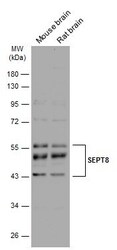
- Western blot analysis of Septin-8 was performed by separating 10 µg of various tissue extracts by 10% SDS-PAGE. Proteins were transferred to a membrane and probed with a Septin-8 Polyclonal Antibody (Product # PA5-31356) at a dilution of 1:4000. The HRP-conjugated anti-rabbit IgG antibody was used to detect the primary antibody.
- Submitted by
- Invitrogen Antibodies (provider)
- Main image
- Experimental details
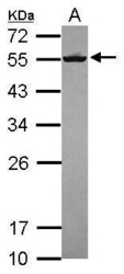
- Western Blot using Septin-8 Polyclonal Antibody (Product # PA5-31356). Sample (30 µg of whole cell lysate). Lane A: HCT116. 12% SDS PAGE. Septin-8 Polyclonal Antibody (Product # PA5-31356) diluted at 1:2,000.
Supportive validation
- Submitted by
- Invitrogen Antibodies (provider)
- Main image
- Experimental details
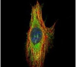
- Immunofluorescent analysis of SEPT8 in methanol-fixed HeLa cells using a SEPT8 polyclonal antibody (Product # PA5-31356) (Green) at a 1:200 dilution. Alpha-tubulin filaments were labeled with Product # PA5-29281 (Red) at a 1:2000.
- Submitted by
- Invitrogen Antibodies (provider)
- Main image
- Experimental details
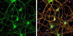
- Immunocytochemistry-Immunofluorescence analysis of Septin-8 was performed in DIV9 rat E18 primary cortical neurons fixed in 4% paraformaldehyde at RT for 15 min. Green: Septin-8 Polyclonal Antibody (Product # PA5-31356) diluted at 1:500. Red: beta Tubulin 3/ Tuj1, stained by beta Tubulin 3/ Tuj1 antibody. Blue: Fluoroshield with DAPI.
Supportive validation
- Submitted by
- Invitrogen Antibodies (provider)
- Main image
- Experimental details
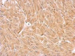
- Septin-8 Polyclonal Antibody detects SEPT8 protein at cytosol on U87 xenograft by immunohistochemical analysis. Sample: Paraffin-embedded U87 xenograft. Septin-8 Polyclonal Antibody (Product # PA5-31356) dilution: 1:500. Antigen Retrieval: EDTA based buffer, pH 8.0, 15 min.
Supportive validation
- Submitted by
- Invitrogen Antibodies (provider)
- Main image
- Experimental details
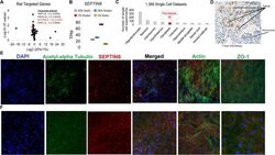
- Figure 5 Data implicating Sept8/ SEPTIN8 in the human tubule hypoxia cell culture model. ( A ) RNAseq values of genes within rat LD block within the RPTEC TERT1 normoxic vs hypoxic conditions. Genes on the right are significant genes. ( B ) TPM values in each of the RNAseq groups for SEPTIN8 in the RPTEC TERT1 cells. ( C ) Expression of SEPTIN8 in single cell datasets (human and mouse). ( D ) Immunohistochemistry from the Human Protein Altas (modified from www.proteinatlas.org/ENSG00000164402-SEPT8/tissue/kidney ) for SEPTIN8 using HPA005665 antibody. Labels are added for tubules and glomerulus. ( E ) Immunofluorescence of SEPTIN8 (red) with acetyl-alpha tubulin, actin, or ZO-1 (green) as labeled. SEPTIN8 localizes with acetyl-alpha tubulin under shear stress with normoxic conditions. ( F ) Under hypoxic conditions, however, SEPTIN8 re-localizes near the actin filaments.
- Submitted by
- Invitrogen Antibodies (provider)
- Main image
- Experimental details
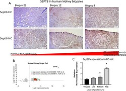
- Figure 6 SEPTIN8/ Sept8 expression increases under conditions of fibrosis or wounding. ( A ) Immunohistochemistry of SEPTIN8 in human biopsies. Results are shown from three separate biopsies with varying levels of tubulointerstitial injury ranging from none (biopsy 22) to moderate (biopsy 12) to severe (biopsy 4). The top panel provides representative low resolution (20X) image of the tubule/interstitial region. The bottom panel provides an independent representative high resolution image (40X) of a single glomerulus. ( B ) Interrogating mouse single-cell databases demonstrates very high levels of Sept8 in response to stimulus or wounding and ( C ) HS rats with high levels of UPE exhibit significantly increased levels of Sept8 relative to HS rats with low to moderate urinary protein levels. rt-qPCR was run in all HS rats that were selected for histological analysis as described in Fig. 1 . * p < 0.05 vs very low, + p < 0.0001 vs all groups.
 Explore
 Validate
 Learn
Learn Western blot