 Explore
 Validate
 Learn
Learn Western blot
Antibody data
- Antibody Data
- Antigen structure
- References [2]
- Comments [0]
- Validations
- Western blot [1]
- Immunohistochemistry [1]
Submit
Validation data
Reference
Comment
Report error
- Product number
- AF739 - Provider product page

- Provider
- R&D Systems
- Product name
- Mouse/Hamster GDF-9 Antibody
- Antibody type
- Polyclonal
- Description
- Antigen Affinity-purified. Detects mouse GDF-9 in direct ELISAs and both mouse and hamster GDF-9 in Western blots. In direct ELISAs, less than 1% cross-reactivity with recombinant mouse (rm) GDF-1, rmGDF-5, rmGDF-6, rmGDF-8, and rmGDF-15 is observed.
- Reactivity
- Mouse, Hamster
- Host
- Goat
- Conjugate
- Unconjugated
- Isotype
- IgG
- Vial size
- 100 ug
- Concentration
- LYOPH
- Storage
- Use a manual defrost freezer and avoid repeated freeze-thaw cycles. 12 months from date of receipt, -20 to -70 °C as supplied. 1 month, 2 to 8 °C under sterile conditions after reconstitution. 6 months, -20 to -70 °C under sterile conditions after reconstitution.
Submitted references GDF9 is transiently expressed in oocytes before follicle formation in the human fetal ovary and is regulated by a novel NOBOX transcript.
Oocyte generation in adult mammalian ovaries by putative germ cells in bone marrow and peripheral blood.
Bayne RA, Kinnell HL, Coutts SM, He J, Childs AJ, Anderson RA
PloS one 2015;10(3):e0119819
PloS one 2015;10(3):e0119819
Oocyte generation in adult mammalian ovaries by putative germ cells in bone marrow and peripheral blood.
Johnson J, Bagley J, Skaznik-Wikiel M, Lee HJ, Adams GB, Niikura Y, Tschudy KS, Tilly JC, Cortes ML, Forkert R, Spitzer T, Iacomini J, Scadden DT, Tilly JL
Cell 2005 Jul 29;122(2):303-15
Cell 2005 Jul 29;122(2):303-15
No comments: Submit comment
Supportive validation
- Submitted by
- R&D Systems (provider)
- Main image
- Experimental details
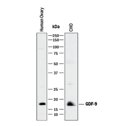
- Detection of Mouse and Hamster GDF-9 by Western Blot. Western blot shows lysates of human ovary tissue and CHO Chinese hamster ovary cell line. PVDF membrane was probed with 1 µg/mL of Goat Anti-Mouse/Hamster GDF-9 Antigen Affinity-purified Polyclonal Antibody (Catalog # AF739) followed by HRP-conjugated Anti-Goat IgG Secondary Antibody (Catalog # HAF017). A specific band was detected for GDF-9 at approximately 18 kDa (as indicated). This experiment was conducted under reducing conditions and using Immunoblot Buffer Group 1.

Supportive validation
- Submitted by
- R&D Systems (provider)
- Main image
- Experimental details
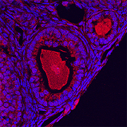
- GDF-9 in Mouse Ovary. GDF-9 was detected in perfusion fixed frozen sections of mouse ovary using Goat Anti-Mouse/Hamster GDF-9 Antigen Affinity-purified Polyclonal Antibody (Catalog # AF739) at 5 µg/mL overnight at 4 °C. Tissue was stained using the NorthernLights™ 557-conjugated Anti-Goat IgG Secondary Antibody (red; Catalog # NL001) and counterstained with DAPI (blue). Specific staining was localized to developing oocytes. View our protocol for Fluorescent IHC Staining of Frozen Tissue Sections.
