 Explore
 Validate
 Learn
Learn Western blot
 ELISA
ELISAAntibody data
- Antibody Data
- Antigen structure
- References [4]
- Comments [0]
- Validations
- Western blot [2]
- Immunocytochemistry [1]
- Immunohistochemistry [1]
- Flow cytometry [1]
Submit
Validation data
Reference
Comment
Report error
- Product number
- ABIN951041 - Provider product page

- Provider
- antibodies-online
- Product name
- anti-Nucleotide-Binding Oligomerization Domain Containing 1 (NOD1) (C-Term) antibody
- Antibody type
- Polyclonal
- Antigen
- Other
- Reactivity
- Human, Mouse
- Host
- Rabbit
- Vial size
- 0.1 mg
Submitted references Association of NOD1 (CARD4) insertion/deletion polymorphism with susceptibility to IBD: a meta-analysis.
Variation at the NFATC2 locus increases the risk of thiazolidinedione-induced edema in the Diabetes REduction Assessment with ramipril and rosiglitazone Medication (DREAM) study.
Helicobacter pylori exploits cholesterol-rich microdomains for induction of NF-kappaB-dependent responses and peptidoglycan delivery in epithelial cells.
Polymorphisms of innate pattern recognition receptors, response to interferon-beta and development of neutralizing antibodies in multiple sclerosis patients.
Lu WG, Zou YF, Feng XL, Yuan FL, Gu YL, Li X, Li CW, Jin C, Li JP
World journal of gastroenterology : WJG 2010 Sep 14;16(34):4348-56
World journal of gastroenterology : WJG 2010 Sep 14;16(34):4348-56
Variation at the NFATC2 locus increases the risk of thiazolidinedione-induced edema in the Diabetes REduction Assessment with ramipril and rosiglitazone Medication (DREAM) study.
Bailey SD, Xie C, Do R, Montpetit A, Diaz R, Mohan V, Keavney B, Yusuf S, Gerstein HC, Engert JC, Anand S, DREAM investigators
Diabetes care 2010 Oct;33(10):2250-3
Diabetes care 2010 Oct;33(10):2250-3
Helicobacter pylori exploits cholesterol-rich microdomains for induction of NF-kappaB-dependent responses and peptidoglycan delivery in epithelial cells.
Hutton ML, Kaparakis-Liaskos M, Turner L, Cardona A, Kwok T, Ferrero RL
Infection and immunity 2010 Nov;78(11):4523-31
Infection and immunity 2010 Nov;78(11):4523-31
Polymorphisms of innate pattern recognition receptors, response to interferon-beta and development of neutralizing antibodies in multiple sclerosis patients.
Enevold C, Oturai AB, Sørensen PS, Ryder LP, Koch-Henriksen N, Bendtzen K
Multiple sclerosis (Houndmills, Basingstoke, England) 2010 Aug;16(8):942-9
Multiple sclerosis (Houndmills, Basingstoke, England) 2010 Aug;16(8):942-9
No comments: Submit comment
Supportive validation
- Submitted by
- antibodies-online (provider)
- Main image
- Experimental details
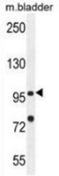
- Western blot analysis in mouse bladder tissue lysates (35ug/lane) using CARD4 / NOD1 antibody Cat.-No. AP52904PU-N (C-term). This demonstrates this antibody detected the NOD1 protein (arrow).

- Submitted by
- antibodies-online (provider)
- Main image
- Experimental details
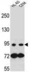
- Western blot analysis in HL-60, CEM cell line lysates (35ug/lane) using CARD4 / NOD1 antibody Cat.-No. AP52904PU-N (C-term). This demonstrates this antibody detected the NOD1 protein (arrow).

Supportive validation
- Submitted by
- antibodies-online (provider)
- Main image

- Experimental details
- Confocal immunofluorescent analysis of NOD1 in HepG2 cell using CARD4 / NOD1 antibody Cat.-No. AP52904PU-N (C-term), followed by Alexa Fluor 488-conjugated goat anti-rabbit lgG (green). DAPI was used to stain the cell nuclear (blue).

Supportive validation
- Submitted by
- antibodies-online (provider)
- Main image

- Experimental details
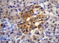
- Immunohistochemistry analysis in human pancreas tissue (Formalin-fixed, Paraffin-embedded) using CARD4 / NOD1 antibody Cat.-No. AP52904PU-N (C-term), followed by peroxidase conjugated secondary antibody and DAB staining. This data demonstrates the use of this antibody for IHC. Clinical relevance has not been evaluated.
Supportive validation
- Submitted by
- antibodies-online (provider)
- Main image

- Experimental details
- Flow cytometric analysis of Jurkat cells (right histogram) compared to a negative control cell (left histogram) using CARD4 / NOD1 antibody Cat.-No. AP52904PU-N (C-term), followed by FITC-conjugated secondary antibodies.
