Explore
 Validate
 Learn
Learn Western blot
Antibody data
- Antibody Data
- Antigen structure
- References [1]
- Comments [0]
- Validations
- Western blot [1]
- Immunocytochemistry [1]
- Immunohistochemistry [2]
- Flow cytometry [1]
Submit
Validation data
Reference
Comment
Report error
- Product number
- 50-9116-42 - Provider product page

- Provider
- Invitrogen Antibodies
- Product name
- Galectin 9 Monoclonal Antibody (9M1-3), eFluor™ 660, eBioscience™
- Antibody type
- Monoclonal
- Antigen
- Other
- Description
- Description: This 9M1-3 monoclonal antibody reacts with human Galectin-9. Galectin-9 is one of fifteen members of the galectin family of glycan-binding proteins. It is expressed on activated T helper cells and Foxp3+ T regulatory cells and plays a critical role in the regulation of immune function. Interaction of Galectin-9 with its ligand, TIM3, induces apoptosis in Th1 and Th17, inhibits Th17 polarization and IFN gamma expression, and drives Treg expansion. Galectin-9 is also thought to contribute to the survival and expansion of some tumors by inhibiting the anti-tumor response and promoting immune evasion. Along with Galectin-1 and Galectin-3, high levels of Galectin-9 expression are associated with poor cancer prognosis, making these potential therapeutic targets.
- Antibody clone number
- 9M1-3
- Concentration
- 5 µL/Test
Submitted references Galectins and their ligands: negative regulators of anti-tumor immunity.
Cedeno-Laurent F, Dimitroff CJ
Glycoconjugate journal 2012 Dec;29(8-9):619-25
Glycoconjugate journal 2012 Dec;29(8-9):619-25
No comments: Submit comment
Supportive validation
- Submitted by
- Invitrogen Antibodies (provider)
- Main image
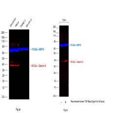
- Experimental details
- Multiplexed fluorescent western blot was performed using Anti-Galectin 9 Monoclonal Antibody (9M1-3), eFluor™ 660, eBioscience™ (Product # 50-9116-42) and a 39 kDa band corresponding to Galectin 9 was observed across the cell lines except in Panc-1 and SK-OV-3. Galectin 9 expression was also upregulated in THP-1 cells upon treatment with Recombinant Human TFG-Beta (2 ng/mL for 24 hours). Whole cell extracts (30 µg lysate) of SH-SY5Y (Lane 1), HeLa (Lane 2), PANC-1 (Lane 3) and SK-OV-3 (Lane 4) as seen in Fig (a) and whole cell extracts (30 µg lysate) of THP-1 (Lane 1) and THP-1 treated with Recombinant Human TFG-Beta (2 ng/mL for 24 hours) (Lane 2) as seen in Fig (b) were electrophoresed using NuPAGE™ 4-12% Bis-Tris Protein Gel (Product # NP0322BOX), 12 well. Resolved proteins were then transferred onto a nitrocellulose membrane (Product # LC2002) by iBlot® 2 Dry Blotting System (Product # IB21001). The blots were probed with the primary antibody (1:500 dilution) and HSP70 Polyclonal Antibody (Product # PA5-28003, 1:4000 dilution). Donkey anti-Rabbit IgG (H+L) Highly Cross-Adsorbed Secondary Antibody, Alexa Fluor™ Plus 800 (Product # A32808, 1:10000 dilution) secondary antibody was used for detection of HSP70. Fluorescent detection was performed using iBright FL1500 (Product # A44115). Relative expression of Galectin 9 was observed to be high in SH-SY5Y and HeLa as compared to PANC-1 and SK-OV-3 cell lines.

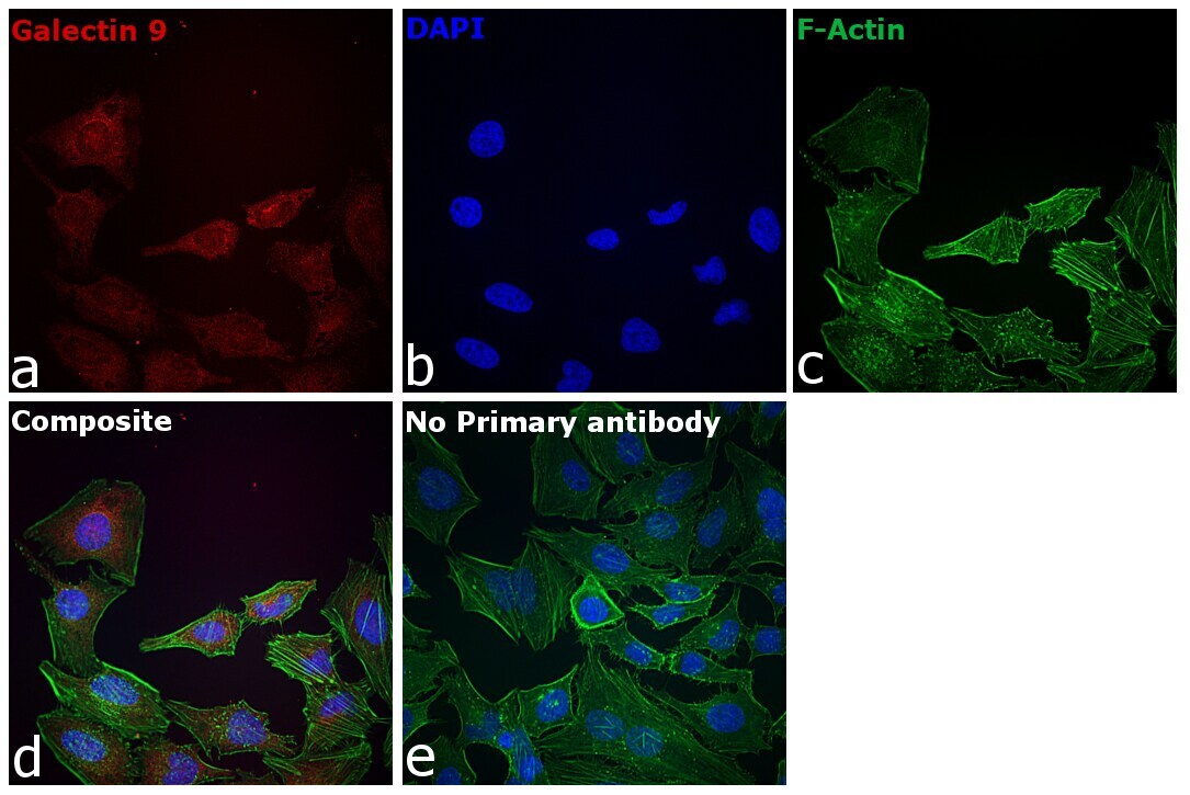
Supportive validation
- Submitted by
- Invitrogen Antibodies (provider)
- Main image
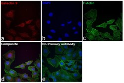
- Experimental details
- Immunofluorescence analysis of Galectin 9 was performed using 70% confluent log phase HeLa cells. The cells were fixed with 4% paraformaldehyde for 10 minutes, permeabilized with 0.1% Triton™ X-100 for 15 minutes, and blocked with 2% BSA for 1 hour at room temperature. The cells were labeled with Galectin 9 Monoclonal Antibody (9M1-3), eFluor™ 660, eBioscience™ (Product # 50-9116-42) at 1:100 dilution in 0.1% BSA, incubated at 4 degree celsius overnight. (Panel a: Red), Nuclei (Panel b:Blue) were stained with Hoechst 33342 (Product # H1399, 1:2000 dilution). F-actin (Panel c: Green) was stained with Alexa Fluor™ 488 Phalloidin (Product # A12379, 1:300 dilution). Panel d represents the merged image showing Nuclear as well as cytoplasmic localization. Panel e represents control cells with no primary antibody to assess background. The images were captured at 40X magnification in CellInsight CX7 LZR High-Content Screening (HCS) Platform (Product # CX7A1110LZR) and externally deconvoluted.
Supportive validation
- Submitted by
- Invitrogen Antibodies (provider)
- Main image

- Experimental details
- Immunohistochemical analysis of Galectin 9 was performed using formalin-fixed paraffin-embedded human colon adenocarcinoma tissue sections. To expose the target protein, heat-induced epitope retrieval was performed on de-paraffinized sections using eBioscience™ IHC Antigen Retrieval Solution - High pH (10X) (Product # 00-4956-58) diluted to 1X solution in water in a decloaking chamber at 110 degree Celsius for 15 minutes. Following antigen retrieval, the sections were blocked with 2% normal goat serum in 1X PBS for 45 minutes at room temperature and then probed with or without Galectin 9 Monoclonal Antibody (9M1-3), eFluor™ 660, eBioscience™ (Product # 50-9116-42) at 1:100 dilution in 0.1% normal goat serum overnight at 4 degree Celsius in a humidified chamber. ReadyProbes™ Tissue Autofluorescence Quenching Kit (Product # R37630) was used to quench autofluorescence from the tissues. Nuclei were stained with DAPI (Product # D1306) and the sections were mounted using ProLong™ Glass Antifade Mountant (Product # P36984). The images were captured on EVOS™ M7000 Imaging System (Product # AMF7000) at 20X magnification and externally deconvoluted.
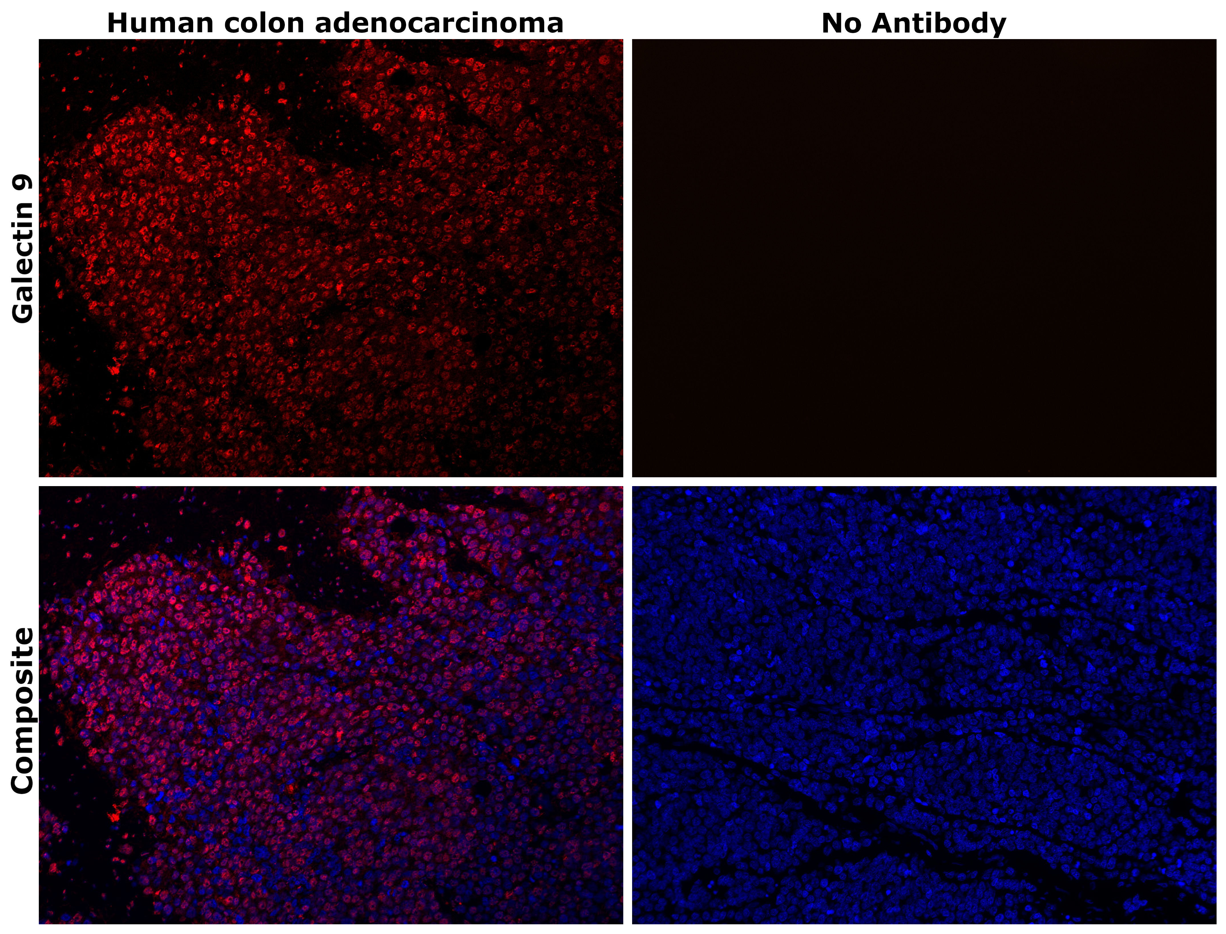
- Submitted by
- Invitrogen Antibodies (provider)
- Main image
- Experimental details
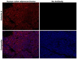
- Immunohistochemical analysis of Galectin 9 was performed using formalin-fixed paraffin-embedded human colon adenocarcinoma tissue sections. To expose the target protein, heat-induced epitope retrieval was performed on de-paraffinized sections using eBioscience™ IHC Antigen Retrieval Solution - High pH (10X) (Product # 00-4956-58) diluted to 1X solution in water in a decloaking chamber at 110 degree Celsius for 15 minutes. Following antigen retrieval, the sections were blocked with 2% normal goat serum in 1X PBS for 45 minutes at room temperature and then probed with or without Galectin 9 Monoclonal Antibody (9M1-3), eFluor™ 660, eBioscience™ (Product # 50-9116-42) at 1:100 dilution in 0.1% normal goat serum overnight at 4 degree Celsius in a humidified chamber. ReadyProbes™ Tissue Autofluorescence Quenching Kit (Product # R37630) was used to quench autofluorescence from the tissues. Nuclei were stained with DAPI (Product # D1306) and the sections were mounted using ProLong™ Glass Antifade Mountant (Product # P36984). The images were captured on EVOS™ M7000 Imaging System (Product # AMF7000) at 20X magnification and externally deconvoluted.

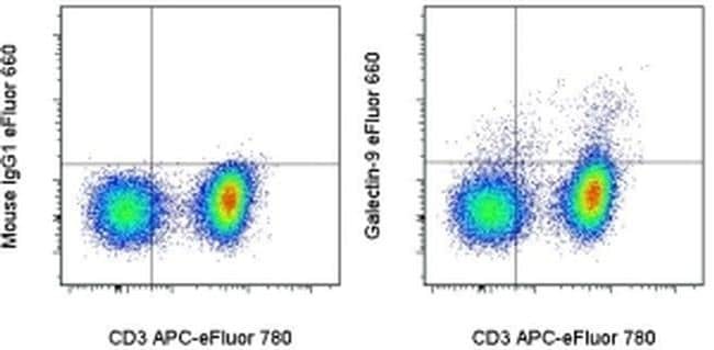
Supportive validation
- Submitted by
- Invitrogen Antibodies (provider)
- Main image
- Experimental details
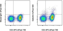
- Normal human peripheral blood cells were stimulated for 3 days with Human IL-2 Recombinant Protein (Product # 14-8029-81). The cells were stained with Anti-Human CD3 APC-eFluor® 780 (Product # 47-0038-42) and Mouse IgG1 K Isotype Control eFluor® 660 (Product # 50-4714-82) (left) or Anti-Human Galectin-9 eFluor® 660 (right). Viable cells in the lymphocyte gate, as determined by Fixable Viability Dye eFluor® 506 (Product # 65-0866-14), were used for analysis.