Explore
 Validate
 Learn
Learn Western blot
Antibody data
- Antibody Data
- Antigen structure
- References [1]
- Comments [0]
- Validations
- Western blot [4]
- Immunocytochemistry [1]
- Immunohistochemistry [1]
Submit
Validation data
Reference
Comment
Report error
- Product number
- GTX108638 - Provider product page

- Provider
- GeneTex
- Proper citation
- GeneTex Cat#GTX108638, RRID:AB_1951937
- Product name
- SMAD3 antibody
- Antibody type
- Polyclonal
- Reactivity
- Human, Mouse, Rat
- Host
- Rabbit
Submitted references BRCA1 regulates microRNA biogenesis via the DROSHA microprocessor complex.
Kawai S, Amano A
The Journal of cell biology 2012 Apr 16;197(2):201-8
The Journal of cell biology 2012 Apr 16;197(2):201-8
No comments: Submit comment
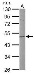
Supportive validation
- Submitted by
- GeneTex (provider)
- Main image

- Experimental details
- Sample (30 ?g of whole cell lysate) A: HeLa 10% SDS PAGE GTX108638 diluted at 1:1000 The HRP-conjugated anti-rabbit IgG antibody (GTX213110-01) was used to detect the primary antibody.

- Submitted by
- GeneTex (provider)
- Main image
- Experimental details
- Sample (50 ug of whole cell lysate) A: Mouse brain 10% SDS PAGE GTX108638 diluted at 1:1000
- Validation comment
- WB

- Submitted by
- GeneTex (provider)
- Main image
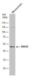
- Experimental details
- Mouse tissue extract (50 ?g) was separated by 10% SDS-PAGE, and the membrane was blotted with SMAD3 antibody (GTX108638) diluted at 1:1000. The HRP-conjugated anti-rabbit IgG antibody (GTX213110-01) was used to detect the primary antibody.

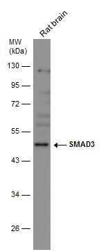
- Submitted by
- GeneTex (provider)
- Main image
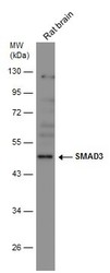
- Experimental details
- Rat tissue extract (50 ?g) was separated by 10% SDS-PAGE, and the membrane was blotted with SMAD3 antibody (GTX108638) diluted at 1:1000. The HRP-conjugated anti-rabbit IgG antibody (GTX213110-01) was used to detect the primary antibody.
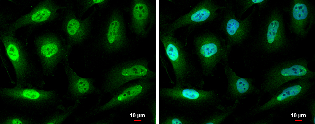
Supportive validation
- Submitted by
- GeneTex (provider)
- Main image

- Experimental details
- SMAD3 antibody detects SMAD3 protein at cytoplasm and nucleus by immunofluorescent analysis.Sample: HeLa cells were fixed in 4% paraformaldehyde at RT for 15 min.Green: SMAD3 protein stained by SMAD3 antibody (GTX108638) diluted at 1:500.Blue: Hoechst 33342 staining.Scale bar = 10 £gm.
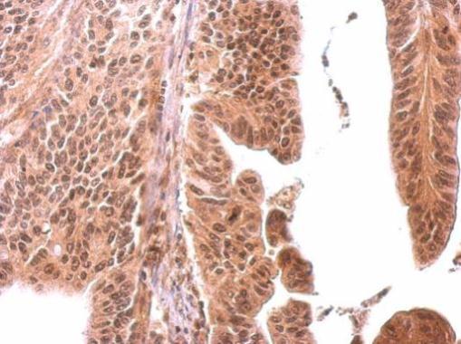
Supportive validation
- Submitted by
- GeneTex (provider)
- Main image

- Experimental details
- SMAD3 antibody detects SMAD3 protein at cytosol and nucleus on human gastric cancer by immunohistochemical analysis. Sample: Paraffin-embedded gastric cancer. SMAD3 antibody (GTX108638) dilution: 1:500.