Explore
 Validate
 Learn
LearnMA5-14939
antibody from Invitrogen Antibodies
Targeting: SMAD3
HsT17436, JV15-2, MADH3
 Western blot Immunocytochemistry
 Immunoprecipitation Flow cytometry Chromatin Immunoprecipitation Other assay
Antibody data
- Antibody Data
- Antigen structure
- References [1]
- Comments [0]
- Validations
- Western blot [2]
- Immunocytochemistry [1]
- Flow cytometry [1]
- Chromatin Immunoprecipitation [1]
- Other assay [1]
Submit
Validation data
Reference
Comment
Report error
- Product number
- MA5-14939 - Provider product page

- Provider
- Invitrogen Antibodies
- Product name
- SMAD3 Monoclonal Antibody (E.980.9)
- Antibody type
- Monoclonal
- Antigen
- Synthetic peptide
- Description
- It is not recommended to aliquot this antibody.
- Reactivity
- Human, Mouse, Rat
- Host
- Rabbit
- Isotype
- IgG
- Antibody clone number
- E.980.9
- Vial size
- 100 µL
- Concentration
- 95.3 µg/mL
- Storage
- -20°C
Submitted references Inhalation of lung spheroid cell secretome and exosomes promotes lung repair in pulmonary fibrosis.
Dinh PC, Paudel D, Brochu H, Popowski KD, Gracieux MC, Cores J, Huang K, Hensley MT, Harrell E, Vandergriff AC, George AK, Barrio RT, Hu S, Allen TA, Blackburn K, Caranasos TG, Peng X, Schnabel LV, Adler KB, Lobo LJ, Goshe MB, Cheng K
Nature communications 2020 Feb 28;11(1):1064
Nature communications 2020 Feb 28;11(1):1064
No comments: Submit comment
Supportive validation
- Submitted by
- Invitrogen Antibodies (provider)
- Main image
- Experimental details
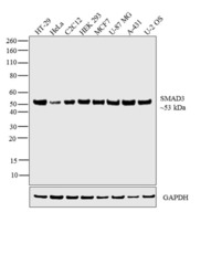
- Western blot analysis was performed on whole cell extracts (30 µg lysate) of HT-29 (Lane 1), HeLa (Lane 2), C2C12 (Lane 3), HEK 293 (Lane 4), MCF7 (Lane 5), U-87 MG (Lane 6), A-431 (Lane 7) and U-2 OS (Lane 8). The blot was probed with Anti-SMAD3 Polyclonal Antibody (Product # MA5-14939, 1:1000 dilution) and detected by chemiluminescence using Goat anti-Rabbit IgG (H+L) Superclonal™ Secondary Antibody, HRP conjugate (Product # A27036, 0.25 µg/mL, 1:4000 dilution). A 53 kDa band corresponding to SMAD3 was observed across the cell lines tested.

- Submitted by
- Invitrogen Antibodies (provider)
- Main image

- Experimental details
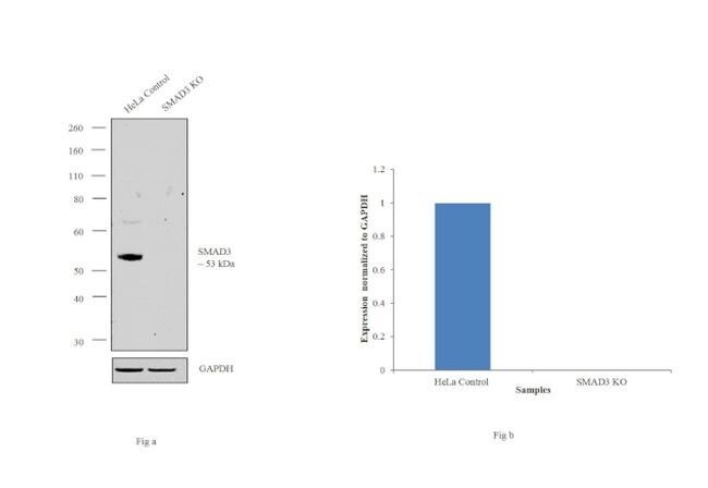
- Western blot analysis of SMAD3 (Fig. a) was performed by loading 20 µg of HeLa Control (lane 1), HeLa SMAD3 knockout (lane 2) whole cell extracts. SMAD3 was detected at 53 kDa using SMAD3 Monoclonal Antibody (E.980.9) (Product # MA5-14939, 1:1000 dilution) and Goat anti-Rabbit IgG (H+L) Superclonal™ Secondary Antibody, HRP conjugate (Product # A27036, 0.25 µg/mL, 1:4000 dilution). Densitometric analysis of this western blot is shown in histogram (Fig. b). Loss of signal in CRISPR mediated knockout (KO) confirms that antibody is specific to SMAD3.
Supportive validation
- Submitted by
- Invitrogen Antibodies (provider)
- Main image
- Experimental details
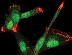
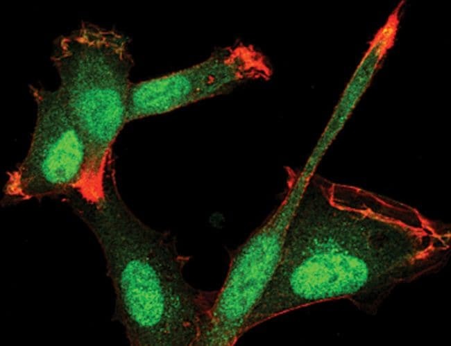
- Immunofluorescent analysis of SMAD3 using a monoclonal antibody (Product # MA5-14939).
Supportive validation
- Submitted by
- Invitrogen Antibodies (provider)
- Main image
- Experimental details
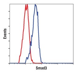
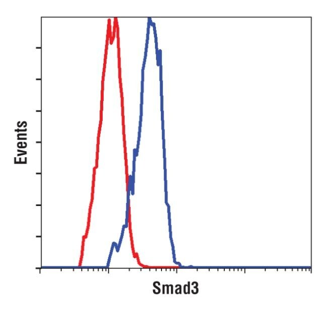
- Flow cytometry analysis of SMAD3 using a monoclonal antibody (Product # MA5-14939).
Supportive validation
- Submitted by
- Invitrogen Antibodies (provider)
- Main image
- Experimental details
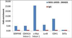
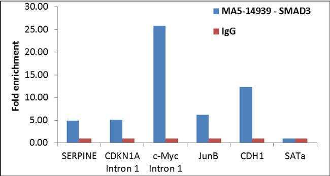
- Enrichment of endogenous SMAD3 protein at specific gene loci using Anti-SMAD3 Antibody: Chromatin Immunoprecipitation (ChIP) was performed using Anti-SMAD3 Mouse Monoclonal Antibody (Product # MA5-14939, 6 µl) on sheared chromatin from 2 million MCF7 cells treated with TGF beta (7 ng/mL for 1 hour) using the MAGnify ChIP system kit (Product # 49-2024). Normal Rabbit IgG was used as a negative IP control. The purified DNA was analyzed by qPCR with PCR primer pairs over SERPINE, CDKN1A Intron 1, c-Myc Intron 1, JunB, CDH1 (active) and SAT alpha (inactive). Data is presented as fold enrichment of the antibody signal versus the negative control IgG using the comparative CT method.
Supportive validation
- Submitted by
- Invitrogen Antibodies (provider)
- Main image

- Experimental details
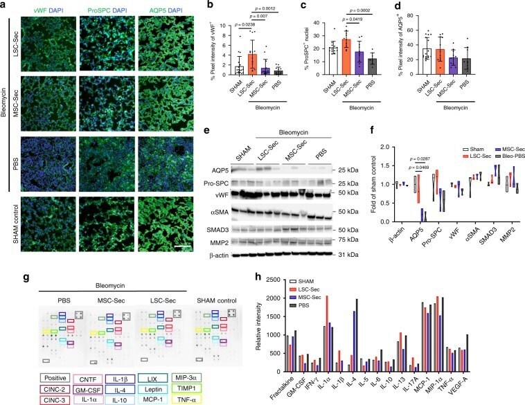
- Fig. 2 LSC-Sec inhalation treatment promotes alveolar repair. a Representative immunostaining of lung sections for von Willebrand Factor (vWF), pro-surfactant protein C (Pro-SPC) and aquaporin 5 (AQP5). Scale bar = 100 mum. b - d Quantification of percent pixel intensity of vWF+ ( b ), percent ProSPC+ nuclei ( c ), and percent pixel intensity of AQP5+ ( d ); each dot represents data from one animal; n = 12 biological independent animals. e - f Immunoblot analysis of aquaporin 5 (AQP5), pro-surfactant protein C (Pro-SPC), von Willebrand Factor (vWF), alpha smooth muscle actin (alphaSMA), SMAD3, matrix metalloproteinase 2 (MMP-2), and beta-actin loading control (B-actin) from lung protein lysate ( e ) with corresponding quantification of protein levels as fold of sham control ( f ); each dot represents data from one animal; n = 3 biological independent animals. g - h Representative cytokine array with quantification of relative intensity ( g ) and corresponding quantification of relative intensity ( h ). Throughout, data are mean +- s.d. P -value as indicated by non-parametric one-way ANOVA.