Explore
 Validate
 Learn
Learn Immunocytochemistry
Antibody data
- Antibody Data
- Antigen structure
- References [1]
- Comments [0]
- Validations
- Immunocytochemistry [1]
- Immunohistochemistry [1]
- Flow cytometry [1]
Submit
Validation data
Reference
Comment
Report error
- Product number
- MAB4038 - Provider product page

- Provider
- R&D Systems
- Product name
- Human Smad3 Antibody
- Antibody type
- Monoclonal
- Description
- Protein A or G purified from hybridoma culture supernatant. Detects human Smad3 in direct ELISAs.
- Reactivity
- Human
- Host
- Rat
- Conjugate
- Unconjugated
- Antigen sequence
P84022- Isotype
- IgG
- Antibody clone number
- 378611
- Vial size
- 100 ug
- Concentration
- LYOPH
- Storage
- Use a manual defrost freezer and avoid repeated freeze-thaw cycles. 12 months from date of receipt, -20 to -70 °C as supplied. 1 month, 2 to 8 °C under sterile conditions after reconstitution. 6 months, -20 to -70 °C under sterile conditions after reconstitution.
Submitted references Transforming growth factor-β and oxidative stress mediate tachycardia-induced cellular remodelling in cultured atrial-derived myocytes.
Yeh YH, Kuo CT, Chan TH, Chang GJ, Qi XY, Tsai F, Nattel S, Chen WJ
Cardiovascular research 2011 Jul 1;91(1):62-70
Cardiovascular research 2011 Jul 1;91(1):62-70
No comments: Submit comment
Supportive validation
- Submitted by
- R&D Systems (provider)
- Main image

- Experimental details
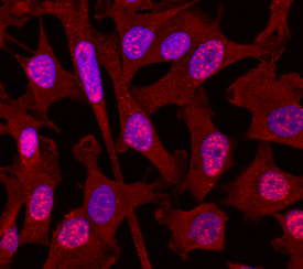
- Smad3 in MDA-MB-231 Human Cell Line. Smad3 was detected in immersion fixed MDA-MB-231 human breast cancer cell line using Rat Anti-Human Smad3 Monoclonal Antibody (Catalog # MAB4038) at 10 µg/mL for 3 hours at room temperature. Cells were stained using the NorthernLights™ 557-conjugated Anti-Rat IgG Secondary Antibody (red; Catalog # NL013) and counterstained with DAPI (blue). Specific staining was localized to cytoplasm and nuclei. View our protocol for Fluorescent ICC Staining of Cells on Coverslips.
Supportive validation
- Submitted by
- R&D Systems (provider)
- Main image
- Experimental details
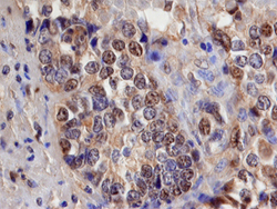
- Smad3 in Human Pancreatic Cancer Tissue. Smad3 was detected in immersion fixed paraffin-embedded sections of human pancreatic cancer tissue using 15 µg/mL Rat Anti-Human Smad3 Monoclonal Antibody (Catalog # MAB4038) overnight at 4 °C. Before incubation with the primary antibody tissue was subjected to heat-induced epitope retrieval using Antigen Retrieval Reagent-Basic (Catalog # CTS013). Tissue was stained with the Anti-Mouse HRP-DAB Cell & Tissue Staining Kit (brown; Catalog # CTS002) and counterstained with hematoxylin (blue). View our protocol for Chromogenic IHC Staining of Paraffin-embedded Tissue Sections.

Supportive validation
- Submitted by
- R&D Systems (provider)
- Main image
- Experimental details
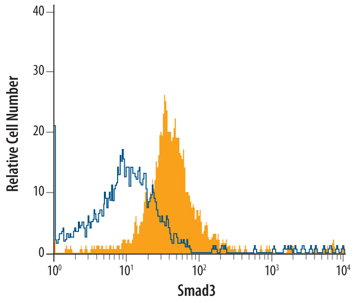
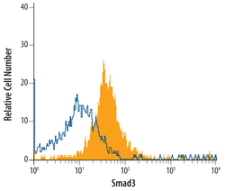
- Detection of Smad3 in PC-3 Human Cell Line by Flow Cytometry. PC-3 human prostate cancer cell line was stained with Rat Anti-Human Smad3 Monoclonal Antibody (Catalog # MAB4038, filled histogram) or isotype control antibody (Catalog # MAB005, open histogram), followed by Allophycocyanin-conjugated Anti-Rat IgG F(ab')2 Secondary Antibody (Catalog # F0113). To facilitate intracellular staining, cells were fixed with paraformaldehyde and permeabilized with methanol.