 Explore
 Validate
 Learn
Learn Immunohistochemistry
Antibody data
- Antibody Data
- Antigen structure
- References [3]
- Comments [0]
- Validations
- Immunohistochemistry [8]
Submit
Validation data
Reference
Comment
Report error
- Product number
- HPA010926 - Provider product page

- Provider
- Atlas Antibodies
- Proper citation
- Atlas Antibodies Cat#HPA010926, RRID:AB_1078449
- Product name
- Anti-ALCAM
- Antibody type
- Polyclonal
- Reactivity
- Human
- Host
- Rabbit
- Conjugate
- Unconjugated
- Antigen sequence
NITLKCLGNGNPPPEEFLFYLPGQPEGIRSSNTYT
LTDVRRNATGDYKCSLIDKKSMIASTAITVHYLDL
SLNPSGEVTRQIGDALPVSCTISASRNATVVWMKD
NIRLRSSPSFSSLHYQDAGNYVCETALQEVEGLKK
R- Isotype
- IgG
- Vial size
- 100 µl
- Storage
- Store at +4°C for short term storage. Long time storage is recommended at -20°C.
Submitted references Elevated ALCAM shedding in colorectal cancer correlates with poor patient outcome.
Membranous expression of activated leukocyte cell adhesion molecule contributes to poor prognosis and malignant phenotypes of non–small-cell lung cancer
Increased expression of ALCAM/CD166 in pancreatic cancer is an independent prognostic marker for poor survival and early tumour relapse
Hansen AG, Freeman TJ, Arnold SA, Starchenko A, Jones-Paris CR, Gilger MA, Washington MK, Fan KH, Shyr Y, Beauchamp RD, Zijlstra A
Cancer research 2013 May 15;73(10):2955-64
Cancer research 2013 May 15;73(10):2955-64
Membranous expression of activated leukocyte cell adhesion molecule contributes to poor prognosis and malignant phenotypes of non–small-cell lung cancer
Ishiguro F, Murakami H, Mizuno T, Fujii M, Kondo Y, Usami N, Taniguchi T, Yokoi K, Osada H, Sekido Y
Journal of Surgical Research 2013 January;179(1):24-32
Journal of Surgical Research 2013 January;179(1):24-32
Increased expression of ALCAM/CD166 in pancreatic cancer is an independent prognostic marker for poor survival and early tumour relapse
Kahlert C, Weber H, Mogler C, Bergmann F, Schirmacher P, Kenngott H, Matterne U, Mollberg N, Rahbari N, Hinz U, Koch M, Aigner M, Weitz J
British Journal of Cancer 2009 July;101(3):457-464
British Journal of Cancer 2009 July;101(3):457-464
No comments: Submit comment
Enhanced validation
Supportive validation
- Submitted by
- Atlas Antibodies (provider)
- Enhanced method
- Orthogonal validation
- Main image

- Experimental details
- Immunohistochemistry analysis in human parathyroid gland and skeletal muscle tissues using HPA010926 antibody. Corresponding ALCAM RNA-seq data are presented for the same tissues.
- Sample type
- HUMAN

Supportive validation
- Submitted by
- Atlas Antibodies (provider)
- Main image

- Experimental details
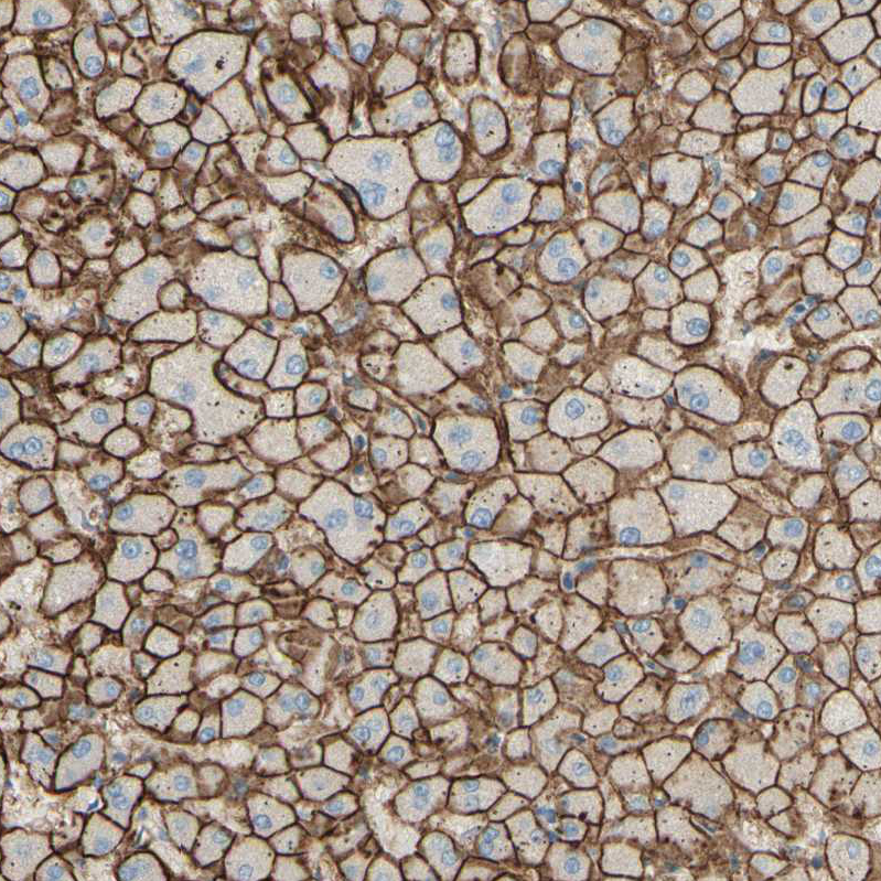
- Immunohistochemical staining of human liver shows distinct membranous positivity in hepatocytes.

- Submitted by
- Atlas Antibodies (provider)
- Main image

- Experimental details
- Immunohistochemical staining of human lung shows strong membranous positivity in pneumocytes.
- Sample type
- HUMAN

- Submitted by
- Atlas Antibodies (provider)
- Main image

- Experimental details
- Immunohistochemical staining of human liver shows strong membranous positivity in hepatocytes.
- Sample type
- HUMAN
- Submitted by
- Atlas Antibodies (provider)
- Main image

- Experimental details
- Immunohistochemical staining of human breast shows strong membranous positivity in glandular cells.
- Sample type
- HUMAN

- Submitted by
- Atlas Antibodies (provider)
- Main image

- Experimental details
- Immunohistochemical staining of human stomach shows strong membranous positivity in glandular cells.
- Sample type
- HUMAN

- Submitted by
- Atlas Antibodies (provider)
- Main image

- Experimental details
- Immunohistochemical staining of human skeletal muscle shows no positivity in myocytes as expected.
- Sample type
- HUMAN

- Submitted by
- Atlas Antibodies (provider)
- Main image

- Experimental details
- Immunohistochemical staining of human parathyroid gland shows strong membranous positivity.
- Sample type
- HUMAN
