Explore
 Validate
 Learn
Learn Western blot
Antibody data
- Antibody Data
- Antigen structure
- References [2]
- Comments [0]
- Validations
- Western blot [6]
- Immunocytochemistry [1]
Submit
Validation data
Reference
Comment
Report error
- Product number
- GTX100416 - Provider product page

- Provider
- GeneTex
- Proper citation
- GeneTex Cat#GTX100416, RRID:AB_1241425
- Product name
- UBE2B antibody
- Antibody type
- Polyclonal
- Reactivity
- Human, Mouse
- Host
- Rabbit
Submitted references Ubiquitin-conjugating enzyme E2 B regulates the ubiquitination of O6-methylguanine-DNA methyltransferase and BCNU sensitivity in human nasopharyngeal carcinoma cells.
Up-regulation of miR-455-5p by the TGF-β-SMAD signalling axis promotes the proliferation of oral squamous cancer cells by targeting UBE2B.
Hsu SH, Chen SH, Kuo CC, Chang JY
Biochemical pharmacology 2018 Dec;158:327-338
Biochemical pharmacology 2018 Dec;158:327-338
Up-regulation of miR-455-5p by the TGF-β-SMAD signalling axis promotes the proliferation of oral squamous cancer cells by targeting UBE2B.
Cheng CM, Shiah SG, Huang CC, Hsiao JR, Chang JY
The Journal of pathology 2016 Sep;240(1):38-49
The Journal of pathology 2016 Sep;240(1):38-49
No comments: Submit comment
Supportive validation
- Submitted by
- GeneTex (provider)
- Main image
- Experimental details
- Sample (30 ug of whole cell lysate) A: NIH-3T3 12% SDS PAGE GTX100416 diluted at 1:1000
- Validation comment
- WB

- Submitted by
- GeneTex (provider)
- Main image
- Experimental details
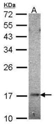
- Sample (30 ug of whole cell lysate) A: Hela 12% SDS PAGE UBE2B antibody GTX100416 diluted at 1:1000
- Validation comment
- WB

- Submitted by
- GeneTex (provider)
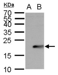
- Main image
- Experimental details
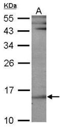
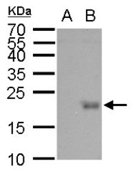
- UBE2B antibody detects UBE2B protein by western blot analysis.A. 30 £gg 293T whole cell lysate/extract B. 30 £gg whole cell lysate/extract of 3xFlag-human UBE2B-transfected 293T cells12 % SDS-PAGEUBE2B antibody (GTX100416) dilution: 1:5000

- Submitted by
- GeneTex (provider)
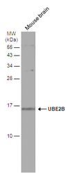
- Main image

- Experimental details
- Mouse tissue extract (50 ?g) was separated by 15% SDS-PAGE, and the membrane was blotted with UBE2B antibody (GTX100416) diluted at 1:500. The signal was developed with Trident ECL plus-Enhanced.

- Submitted by
- GeneTex (provider)
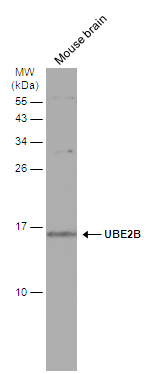
- Main image
- Experimental details
- Mouse tissue extract (50 ?g) was separated by 15% SDS-PAGE, and the membrane was blotted with UBE2B antibody (GTX100416) diluted at 1:1000. The HRP-conjugated anti-rabbit IgG antibody (GTX213110-01) was used to detect the primary antibody.
- Submitted by
- GeneTex (provider)
- Main image
- Experimental details
- UBE2B antibody detects UBE2B protein by Western blot analysis.A. 30 £gg 293T whole cell lysate/extract B. 30 £gg whole cell lysate/extract of 3xFlag-human UBE2B-transfected 293T cells12 % SDS-PAGEUBE2B antibody (GTX100416) dilution: 1:5000

Supportive validation
- Submitted by
- GeneTex (provider)
- Main image

- Experimental details
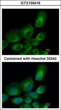
- Immunofluorescence analysis of paraformaldehyde-fixed A431, using UBE2B(GTX100416) antibody at 1:500 dilution.