 Explore
 Validate
 Learn
Learn Western blot
Antibody data
- Antibody Data
- Antigen structure
- References [0]
- Comments [0]
- Validations
- Western blot [3]
- Immunocytochemistry [1]
- Immunohistochemistry [1]
Submit
Validation data
Reference
Comment
Report error
- Product number
- PA5-27111 - Provider product page

- Provider
- Invitrogen Antibodies
- Product name
- TDP1 Polyclonal Antibody
- Antibody type
- Polyclonal
- Antigen
- Recombinant protein fragment
- Reactivity
- Human
- Host
- Rabbit
- Isotype
- IgG
- Vial size
- 100 µL
- Concentration
- 1 mg/mL
- Storage
- Store at 4°C short term. For long term storage, store at -20°C, avoiding freeze/thaw cycles.
No comments: Submit comment
Supportive validation
- Submitted by
- Invitrogen Antibodies (provider)
- Main image
- Experimental details
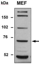
- Western blot analysis of TDP1 was performed by loading whole cell lysate in 1X SDS sample buffer with 2-ME from 5 x 10⁵ mouse embryonic fibroblasts (MEF). Samples were loaded onto a 4-12 % Bis-Tris polyacrylamide gel (Product # WG1402BOX). Proteins were transferred to nitrocellulose membrane with wet/tank transfer. Membrane was blocked in 5% milk/TBST. TDP1 was detected at approximately 60 kDa using a polyclonal anti-TDP1 antibody (Product # PA5-27111) at a dilution of 1:1000 in 5% milk/TBST at 4°C overnight, followed by a secondary antibody HRP-anti-rabbit at a dilution of 1:5000 at room temperature for 1 hour. Chemiluminescent detection was performed using Pierce ECL Western Blotting Substrate (Product # PI-32209). Data courtesy of Antibody Data Exchange Program.

- Submitted by
- Invitrogen Antibodies (provider)
- Main image
- Experimental details
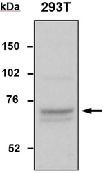
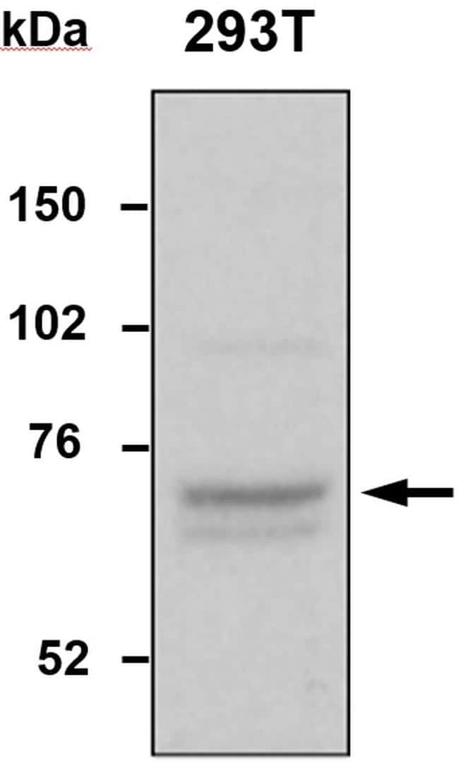
- Western blot analysis of TDP1 was performed by loading whole cell lysate in 1X SDS sample buffer with 2-ME from 106 293T cells. Samples were loaded onto a 4-12 % Bis-Tris polyacrylamide gel (Product # WG1402BOX). Proteins were transferred to nitrocellulose membrane with wet/tank transfer. Membrane was blocked in 5% milk/TBST. TDP1 was detected at approximately 60 kDa using a polyclonal anti-TDP1 antibody (Product # PA5-27111) at a dilution of 1:1000 in 5% milk/TBST at 4°C overnight, followed by a secondary antibody HRP-anti-rabbit at a dilution of 1:5000 at room temperature for 1 hour. Chemiluminescent detection was performed using Pierce ECL Western Blotting Substrate (Product # PI-32209). Data courtesy of Antibody Data Exchange Program.
- Submitted by
- Invitrogen Antibodies (provider)
- Main image

- Experimental details
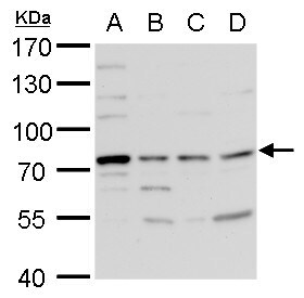
- TDP1 Polyclonal Antibody detects TDP1 protein by western blot analysis. A. 30 µg 293T whole cell lysate/extract. B. 30 µg A431 whole cell lysate/extract. C. 30 µg HeLa whole cell lysate/extract. D. 30 µg HepG2 whole cell lysate/extract.7.5 % SDS-PAGE. TDP1 Polyclonal Antibody (Product # PA5-27111) dilution: 1:5,000.
Supportive validation
- Submitted by
- Invitrogen Antibodies (provider)
- Main image

- Experimental details
- Immunofluorescent analysis of TDP1 in methanol-fixed A431 cells using a TDP1 polyclonal antibody (Product # PA5-27111) at a 1:200 dilution.

Supportive validation
- Submitted by
- Invitrogen Antibodies (provider)
- Main image

- Experimental details
- Immunohistochemical analysis of paraffin-embedded human gastric, using TDP1 (Product # PA5-27111) antibody at 1:100 dilution. Antigen Retrieval: EDTA based buffer, pH 8.0, 15 min.
