Explore
 Validate
 Learn
Learn Western blot
 Immunohistochemistry
Antibody data
- Antibody Data
- Antigen structure
- References [0]
- Comments [0]
- Validations
- Western blot [3]
- Immunocytochemistry [2]
- Immunohistochemistry [7]
Submit
Validation data
Reference
Comment
Report error
- Product number
- HPA023309 - Provider product page

- Provider
- Atlas Antibodies
- Proper citation
- Atlas Antibodies Cat#HPA023309, RRID:AB_10600093
- Product name
- Anti-SEPT7
- Antibody type
- Polyclonal
- Reactivity
- Human, Mouse, Rat
- Host
- Rabbit
- Conjugate
- Unconjugated
- Antigen sequence
MSVSARSAAAEERSVNSSTMVAQQKNLEGYVGFAN
LPNQVYRKSVKRGFEFTLMVVGESG- Isotype
- IgG
- Vial size
- 100 µl
- Storage
- Store at +4°C for short term storage. Long time storage is recommended at -20°C.
No comments: Submit comment
Supportive validation
Supportive validation
- Submitted by
- Atlas Antibodies (provider)
- Enhanced method
- Independent antibody validation
- Main image

- Experimental details
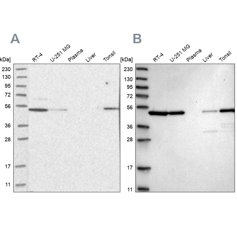
- Western blot analysis using Anti-SEPT7 antibody HPA023309 (A) shows similar pattern to independent antibody HPA029524 (B).
Supportive validation
- Submitted by
- Atlas Antibodies (provider)
- Main image

- Experimental details
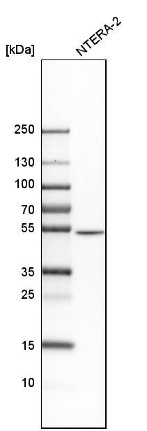
- Western blot analysis in human cell line NTERA-2.
- Submitted by
- Atlas Antibodies (provider)
- Main image

- Experimental details
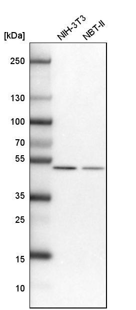
- Western blot analysis in mouse cell line NIH-3T3 and rat cell line NBT-II.
Supportive validation
- Submitted by
- Atlas Antibodies (provider)
- Main image

- Experimental details
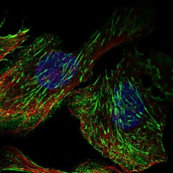
- Immunofluorescent staining of human cell line A-431 shows localization to actin filaments & microtubules.

- Submitted by
- Atlas Antibodies (provider)
- Main image
- Experimental details
- Immunofluorescent staining of human cell line U-2 OS shows localization to actin filaments & midbody.
- Sample type
- HUMAN

Enhanced validation
Enhanced validation
Supportive validation
- Submitted by
- Atlas Antibodies (provider)
- Enhanced method
- Orthogonal validation
- Main image
- Experimental details
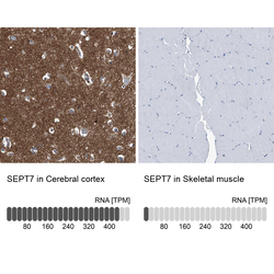
- Immunohistochemistry analysis in human cerebral cortex and skeletal muscle tissues using HPA023309 antibody. Corresponding SEPT7 RNA-seq data are presented for the same tissues.
- Sample type
- HUMAN

Enhanced validation
- Submitted by
- Atlas Antibodies (provider)
- Enhanced method
- Independent antibody validation
- Main image
- Experimental details
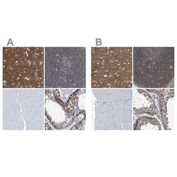
- Immunohistochemical staining of human cerebral cortex, lymph node, skeletal muscle and testis using Anti-SEPT7 antibody HPA023309 (A) shows similar protein distribution across tissues to independent antibody HPA029524 (B).

Supportive validation
- Submitted by
- Atlas Antibodies (provider)
- Main image

- Experimental details
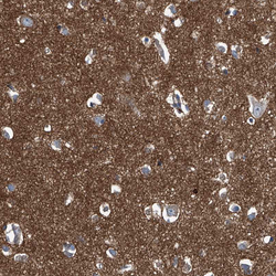
- Immunohistochemical staining of human cerebellum shows moderate cytoplasmic positivity in Purkinje cells.

- Submitted by
- Atlas Antibodies (provider)
- Main image
- Experimental details
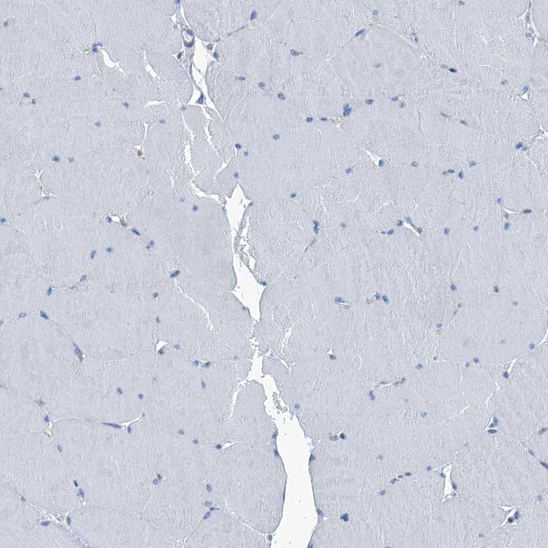
- Immunohistochemical staining of human cerebral cortex shows positivity in neuropil.
- Sample type
- HUMAN

- Submitted by
- Atlas Antibodies (provider)
- Main image
- Experimental details
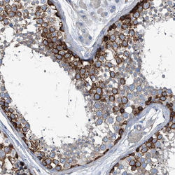
- Immunohistochemical staining of human testis shows positivit in cells in seminiferous ducts.
- Sample type
- HUMAN

- Submitted by
- Atlas Antibodies (provider)
- Main image

- Experimental details
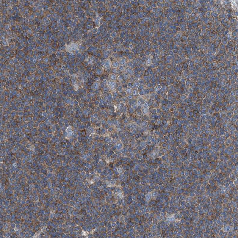
- Immunohistochemical staining of human lymph node shows moderate cytoplasmic positivity in lymphoid cells.
- Sample type
- HUMAN
- Submitted by
- Atlas Antibodies (provider)
- Main image
- Experimental details
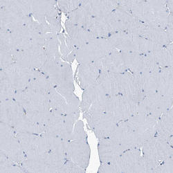
- Immunohistochemical staining of human skeletal muscle shows no positivity in myocytes as expected.
- Sample type
- HUMAN