Explore
 Validate
 Learn
Learn Western blot
Antibody data
- Antibody Data
- Antigen structure
- References [2]
- Comments [0]
- Validations
- Western blot [4]
- Immunoprecipitation [1]
Submit
Validation data
Reference
Comment
Report error
- Product number
- NB100-511 - Provider product page

- Provider
- Novus Biologicals
- Proper citation
- Novus Cat#NB100-511, RRID:AB_2096744
- Product name
- Rabbit Polyclonal EIF3C Antibody
- Antibody type
- Polyclonal
- Description
- Immunogen affinity purified.
- Reactivity
- Human, Mouse
- Host
- Rabbit
- Isotype
- IgG
- Vial size
- 100 ul
- Concentration
- 1.0 mg/ml
- Storage
- Store at 4C. Do not freeze.
Submitted references Differential Sensitivity of Target Genes to Translational Repression by miR-17~92.
EJC core component MLN51 interacts with eIF3 and activates translation.
Jin HY, Oda H, Chen P, Yang C, Zhou X, Kang SG, Valentine E, Kefauver JM, Liao L, Zhang Y, Gonzalez-Martin A, Shepherd J, Morgan GJ, Mondala TS, Head SR, Kim PH, Xiao N, Fu G, Liu WH, Han J, Williamson JR, Xiao C
PLoS genetics 2017 Feb;13(2):e1006623
PLoS genetics 2017 Feb;13(2):e1006623
EJC core component MLN51 interacts with eIF3 and activates translation.
Chazal PE, Daguenet E, Wendling C, Ulryck N, Tomasetto C, Sargueil B, Le Hir H
Proceedings of the National Academy of Sciences of the United States of America 2013 Apr 9;110(15):5903-8
Proceedings of the National Academy of Sciences of the United States of America 2013 Apr 9;110(15):5903-8
No comments: Submit comment
Supportive validation
- Submitted by
- Novus Biologicals (provider)
- Main image
- Experimental details
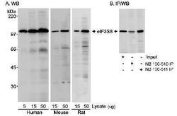
- Western Blot: EIF3C Antibody [NB100-511] - Detection of Human, Mouse and Rat eIF3S8 using NB 100-511. Samples: A. Whole cell lysate (5, 15 or 50 ug for WB) from HEK293 (human), C2C12 (mouse) and L6 (rat) cells. B. Whole cell lysate (12.5 ug for Input and 1 mg for IP) from HEK293 cells. Immunoprecipitation was also performed using NB 100-510 rabbit anti-eIF3S8 antibody at 5 ug/mg lysate. Chemiluminescence with exposure times of 2-4 minutes.

- Submitted by
- Novus Biologicals (provider)
- Main image

- Experimental details
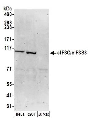
- Western Blot: EIF3C Antibody [NB100-511] - Detection of Human eIF3C/eIF3S8 by Western Blot. Samples: Whole cell lysate (50 ug) from HeLa, 293T, and Jurkat cells prepared using NETN lysis buffer. Antibodies: Affinity purified rabbit anti-eIF3C/eIF3S8 antibody NB100-511 used for WB at 0.1 ug/ml. Detection: Chemiluminescence with an exposure time of 3 minutes.
- Submitted by
- Novus Biologicals (provider)
- Main image

- Experimental details
- Western Blot: EIF3C Antibody [NB100-511] - eIF3C expression in naive and activated (overnight) murine B cells. This image was submitted via customer Review.

- Submitted by
- Novus Biologicals (provider)
- Main image

- Experimental details
- Western Blot: EIF3C Antibody [NB100-511] - eIF3C expression in naive and activated (overnight) murine B cells. Image submitted by a verified customer review.

Supportive validation
- Submitted by
- Novus Biologicals (provider)
- Main image

- Experimental details
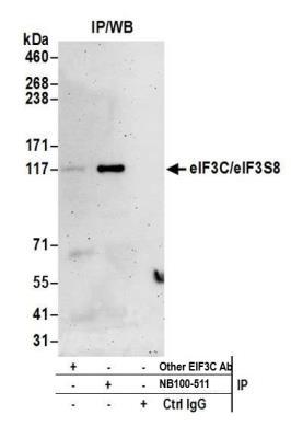
- Immunoprecipitation: EIF3C Antibody [NB100-511] - Detection of human eIF3C/eIF3S8 by western blot of immunoprecipitates. Samples: Whole cell lysate (0.5 or 1.0 mg per IP reaction; 20% of IP loaded) from HEK293T cells prepared using NETN lysis buffer. Antibodies: Affinity purified rabbit anti-eIF3C/eIF3S8 antibody NB100-511 used for IP at 6 ug per reaction. eIF3C/eIF3S8 was inefficiently immunoprecipitated by rabbit anti-eIF3C/eIF3S8 antibody from Company B. For blotting immunoprecipitated eIF3C/eIF3S8, NB100-511 was used at 1 ug/ml. Detection: Chemiluminescence with an exposure time of 3 minutes.