 Explore
 Validate
 Learn
LearnAF6805
antibody from R&D Systems
Targeting: ITGB1BP1
ICAP-1A, ICAP-1alpha, ICAP-1B, ICAP1, ICAP1A, ICAP1B
 Western blot
Antibody data
- Antibody Data
- Antigen structure
- References [1]
- Comments [0]
- Validations
- Western blot [1]
- Immunohistochemistry [1]
Submit
Validation data
Reference
Comment
Report error
- Product number
- AF6805 - Provider product page

- Provider
- R&D Systems
- Product name
- Human ICAP-1 Antibody
- Antibody type
- Polyclonal
- Description
- Immunogen affinity purified. Detects human ICAP-1 in direct ELISAs and Western blots.
- Reactivity
- Human
- Host
- Sheep
- Conjugate
- Unconjugated
- Antigen sequence
O14713- Isotype
- IgG
- Vial size
- 100 ug
- Concentration
- LYOPH
- Storage
- Use a manual defrost freezer and avoid repeated freeze-thaw cycles. 12 months from date of receipt, -20 to -70 °C as supplied. 1 month, 2 to 8 °C under sterile conditions after reconstitution. 6 months, -20 to -70 °C under sterile conditions after reconstitution.
Submitted references Nuclear Localization of Integrin Cytoplasmic Domain-associated Protein-1 (ICAP1) Influences β1 Integrin Activation and Recruits Krev/Interaction Trapped-1 (KRIT1) to the Nucleus.
Draheim KM, Huet-Calderwood C, Simon B, Calderwood DA
The Journal of biological chemistry 2017 Feb 3;292(5):1884-1898
The Journal of biological chemistry 2017 Feb 3;292(5):1884-1898
No comments: Submit comment
Supportive validation
- Submitted by
- R&D Systems (provider)
- Main image
- Experimental details
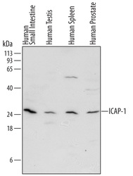
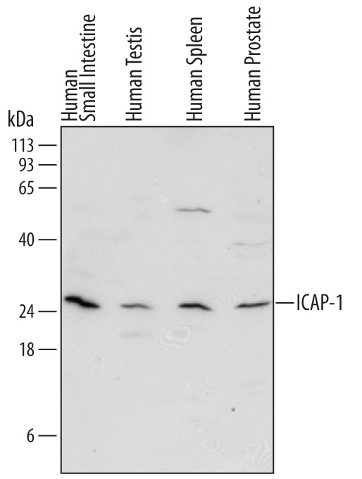
- Detection of Human ICAP-1 by Western Blot. Western blot shows lysates of human small intestine tissue, human testis tissue, human spleen tissue, and human prostate tissue. PVDF Membrane was probed with 2 µg/mL of Sheep Anti-Human ICAP-1 Antigen Affinity-purified Polyclonal Antibody (Catalog # AF6805) followed by HRP-conjugated Anti-Sheep IgG Secondary Antibody (Catalog # HAF016). A specific band was detected for ICAP-1 at approximately 25 kDa (as indicated). This experiment was conducted under reducing conditions and using Immunoblot Buffer Group 1.
Supportive validation
- Submitted by
- R&D Systems (provider)
- Main image
- Experimental details
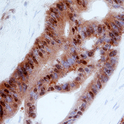
- ICAP-1 in Human Colon Cancer Tissue. ICAP-1 was detected in immersion fixed paraffin-embedded sections of human colon cancer tissue using Sheep Anti-Human ICAP-1 Antigen Affinity-purified Polyclonal Antibody (Catalog # AF6805) at 3 µg/mL overnight at 4 °C. Before incubation with the primary antibody, tissue was subjected to heat-induced epitope retrieval using Antigen Retrieval Reagent-Basic (Catalog # CTS013). Tissue was stained using the Anti-Sheep HRP-DAB Cell & Tissue Staining Kit (brown; Catalog # CTS019) and counterstained with hematoxylin (blue). Specific staining was localized to nuclei in epithelial cells. View our protocol for Chromogenic IHC Staining of Paraffin-embedded Tissue Sections.
