 Explore
 Validate
 Learn
Learn Immunocytochemistry
Antibody data
- Antibody Data
- Antigen structure
- References [0]
- Comments [0]
- Validations
- Immunocytochemistry [1]
- Immunohistochemistry [2]
- Flow cytometry [1]
Submit
Validation data
Reference
Comment
Report error
- Product number
- MA5-29046 - Provider product page

- Provider
- Invitrogen Antibodies
- Product name
- ASGR2 Recombinant Rabbit Monoclonal Antibody (2)
- Antibody type
- Monoclonal
- Antigen
- Recombinant full-length protein
- Description
- This product is preservative free. It is recommended to add sodium azide to avoid contamination (final concentration 0.05%-0.1%).
- Antibody clone number
- 2
- Concentration
- 1.0 mg/mL
No comments: Submit comment
Supportive validation
- Submitted by
- Invitrogen Antibodies (provider)
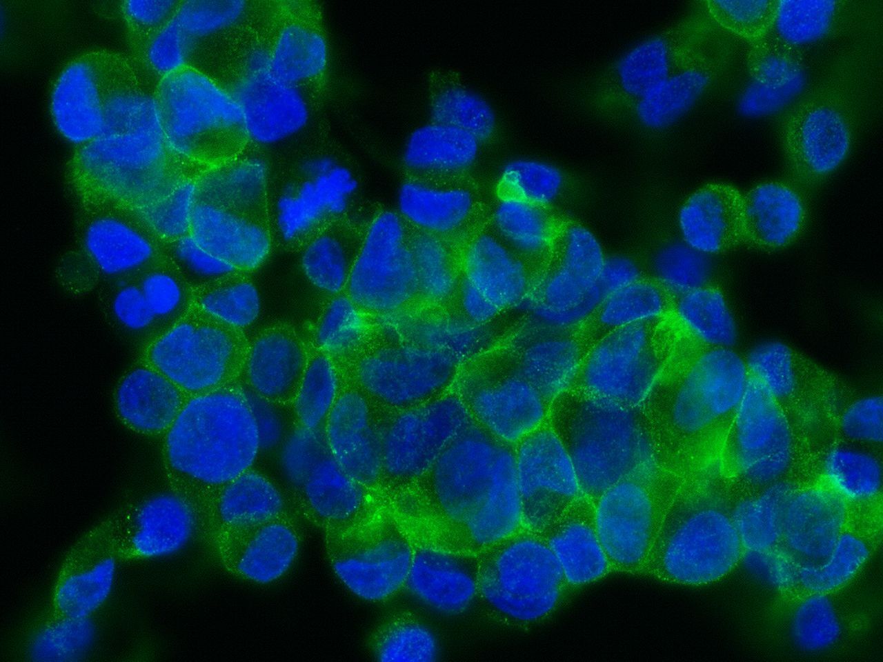
- Main image

- Experimental details
- Immunofluorescence staining of ASGR2 in HepG2 cells. Cells were fixed with 4% PFA, permeabilzed with 0.3% Triton X-100 in PBS, blocked with 10% serum, and incubated with ASGR2 Recombinant Rabbit Monoclonal Antibody (2) (Product # MA5-29046, 1:60) at 4°C overnight. Then cells were stained with the Alexa Fluor® 488-conjugated Goat Anti-rabbit IgG secondary antibody (green) and counterstained with DAPI (blue).
Supportive validation
- Submitted by
- Invitrogen Antibodies (provider)
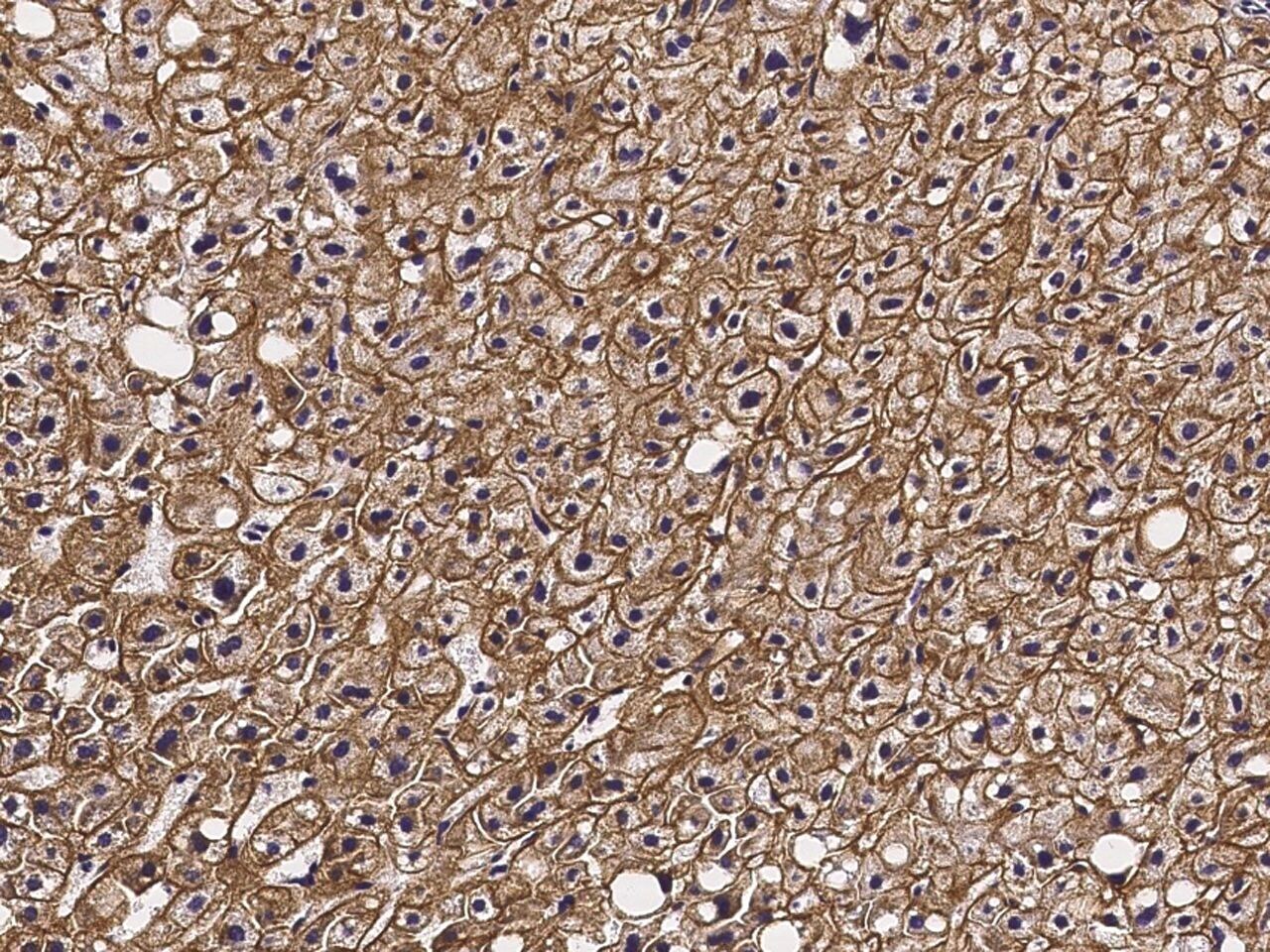
- Main image

- Experimental details
- Immunohistochemical staining of human ASGR2 in human cirrhosis with ASGR2 Recombinant Rabbit Monoclonal Antibody (2) (Product # MA5-29046, 1:200, formalin-fixed paraffin embedded sections).
- Submitted by
- Invitrogen Antibodies (provider)
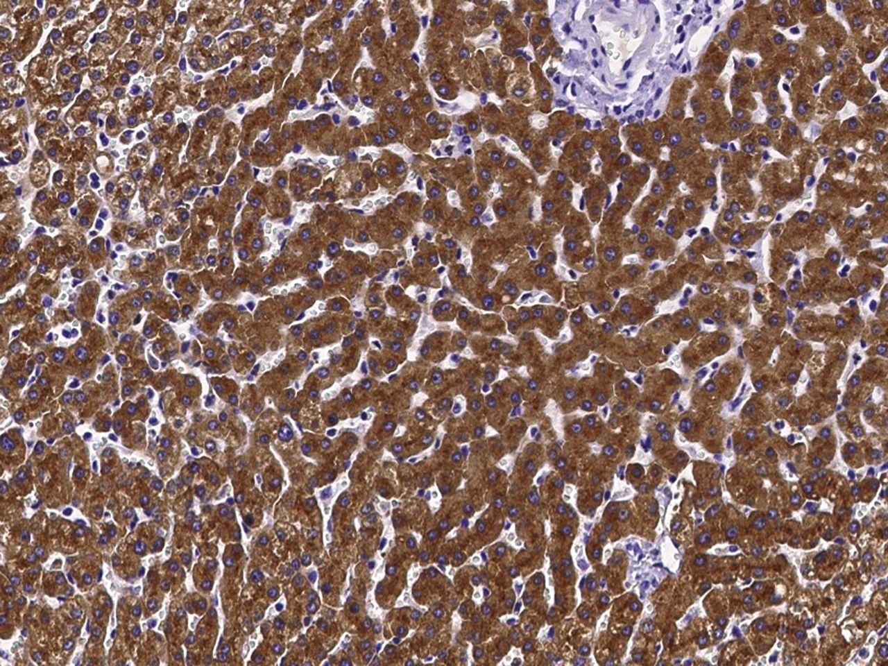
- Main image

- Experimental details
- Immunohistochemical staining of human ASGR2 in human liver with ASGR2 Recombinant Rabbit Monoclonal Antibody (2) (Product # MA5-29046, 1:200, formalin-fixed paraffin embedded sections).
Supportive validation
- Submitted by
- Invitrogen Antibodies (provider)
- Main image

- Experimental details
- Flow cytometric analysis of Human ASGR2 expression on HepG2 cells. Cells were stained with ASGR2 Recombinant Rabbit Monoclonal Antibody (2) (Product # MA5-29046), then a FITC-conjugated Secondary antibody. The fluorescence histograms were derived from gated events with the forward and side light-scatter characteristics of intact cells.
