 Explore
 Validate
 Learn
Learn Western blot
 Immunohistochemistry
Antibody data
- Antibody Data
- Antigen structure
- References [0]
- Comments [0]
- Validations
- Western blot [2]
- Immunocytochemistry [2]
- Immunohistochemistry [13]
Submit
Validation data
Reference
Comment
Report error
- Product number
- HPA014907 - Provider product page

- Provider
- Atlas Antibodies
- Proper citation
- Atlas Antibodies Cat#HPA014907, RRID:AB_1855166
- Product name
- Anti-PDZK1IP1
- Antibody type
- Polyclonal
- Description
- Polyclonal Antibody against Human PDZK1IP1, Gene description: PDZK1 interacting protein 1, Alternative Gene Names: DD96, MAP17, SPAP, Validated applications: ICC, IHC, WB, Uniprot ID: Q13113, Storage: Store at +4°C for short term storage. Long time storage is recommended at -20°C.
- Reactivity
- Human
- Host
- Rabbit
- Conjugate
- Unconjugated
- Isotype
- IgG
- Vial size
- 100 µl
- Storage
- Store at +4°C for short term storage. Long time storage is recommended at -20°C.
No comments: Submit comment
Supportive validation
- Submitted by
- Atlas Antibodies (provider)
- Enhanced method
- Recombinant expression validation
- Main image

- Experimental details
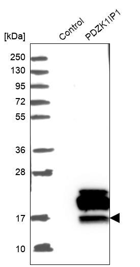
- Western blot analysis in control (vector only transfected HEK293T lysate) and PDZK1IP1 over-expression lysate (Co-expressed with a C-terminal myc-DDK tag (~3.1 kDa) in mammalian HEK293T cells, LY417088).

- Submitted by
- Atlas Antibodies (provider)
- Main image

- Experimental details
- Western blot analysis in control (vector only transfected HEK293T lysate) and PDZK1IP1 over-expression lysate (Co-expressed with a C-terminal myc-DDK tag (~3.1 kDa) in mammalian HEK293T cells, LY417088).
- Sample type
- Human
- Protocol
- Protocol
Supportive validation
- Submitted by
- Atlas Antibodies (provider)
- Main image

- Experimental details
- Immunofluorescent staining of human cell line RPTEC TERT1 shows localization to nuclear speckles & cytosol.
- Sample type
- HUMAN

- Submitted by
- Atlas Antibodies (provider)
- Main image

- Experimental details
- Immunofluorescent staining of human cell line RPTEC TERT1 shows localization to nuclear speckles & cytosol.
- Sample type
- Human
- Protocol
- Protocol

Enhanced validation
Enhanced validation
Supportive validation
- Submitted by
- Atlas Antibodies (provider)
- Enhanced method
- Orthogonal validation
- Main image

- Experimental details
- Immunohistochemistry analysis in human kidney and endometrium tissues using HPA014907 antibody. Corresponding PDZK1IP1 RNA-seq data are presented for the same tissues.
- Sample type
- HUMAN

Enhanced validation
- Submitted by
- Atlas Antibodies (provider)
- Enhanced method
- Orthogonal validation
- Main image

- Experimental details
- Immunohistochemistry analysis in human kidney and endometrium tissues using HPA014907 antibody. Corresponding PDZK1IP1 RNA-seq data are presented for the same tissues.
- Sample type
- Human
- Protocol
- Protocol

Supportive validation
- Submitted by
- Atlas Antibodies (provider)
- Main image

- Experimental details
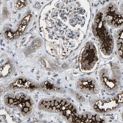
- Immunohistochemical staining of human kidney shows strong luminal membranous positivity in cells in tubules.

- Submitted by
- Atlas Antibodies (provider)
- Main image
- Experimental details
- Immunohistochemical staining of human kidney shows high expression.
- Sample type
- HUMAN

- Submitted by
- Atlas Antibodies (provider)
- Main image
- Experimental details
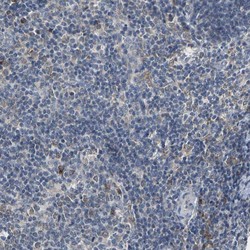
- Immunohistochemical staining of human lymph node shows low expression as expected.
- Sample type
- HUMAN

- Submitted by
- Atlas Antibodies (provider)
- Main image

- Experimental details
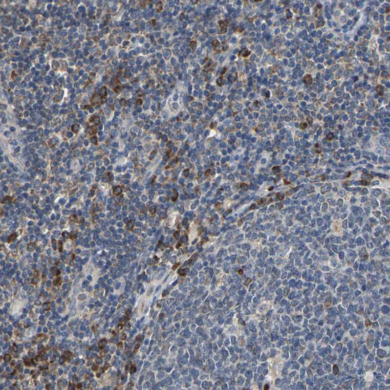
- Immunohistochemical staining of human lymphoid tissues shows moderate cytoplasmic positivity in non-germinal center cells.
- Sample type
- HUMAN
- Submitted by
- Atlas Antibodies (provider)
- Main image
- Experimental details
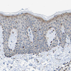
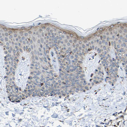
- Immunohistochemical staining of human skin shows weak membranous and cytoplasmic positivity in superficial layer of squamous epithelial cells.
- Sample type
- HUMAN

- Submitted by
- Atlas Antibodies (provider)
- Main image

- Experimental details
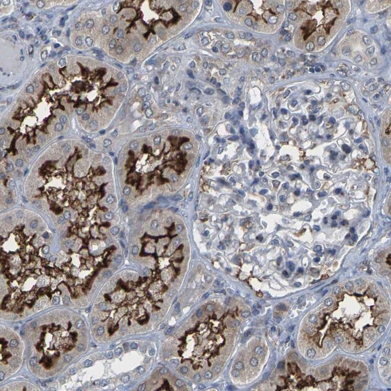
- Immunohistochemical staining of human kidney shows strong strong positivity in apical membrane in cells in tubules.
- Sample type
- HUMAN
- Submitted by
- Atlas Antibodies (provider)
- Main image

- Experimental details
- Immunohistochemical staining of human endometrium shows no positivity in glandular cells as expected.
- Sample type
- HUMAN

- Submitted by
- Atlas Antibodies (provider)
- Main image

- Experimental details
- Immunohistochemical staining of human kidney shows strong strong positivity in apical membrane in cells in tubules.
- Sample type
- Human
- Protocol
- Protocol

- Submitted by
- Atlas Antibodies (provider)
- Main image
- Experimental details
- Immunohistochemical staining of human skin shows weak membranous and cytoplasmic positivity in superficial layer of squamous epithelial cells.
- Sample type
- Human
- Protocol
- Protocol

- Submitted by
- Atlas Antibodies (provider)
- Main image

- Experimental details
- Immunohistochemical staining of human lymphoid tissues shows moderate cytoplasmic positivity in non-germinal center cells.
- Sample type
- Human
- Protocol
- Protocol

- Submitted by
- Atlas Antibodies (provider)
- Main image

- Experimental details
- Immunohistochemical staining of human endometrium shows no positivity in glandular cells as expected.
- Sample type
- Human
- Protocol
- Protocol
