 Explore
 Validate
 Learn
LearnHPA030443
antibody from Atlas Antibodies
Targeting: PARD3
ASIP, Baz, Bazooka, PAR3, PARD3A, PPP1R118
 Western blot
Antibody data
- Antibody Data
- Antigen structure
- References [0]
- Comments [0]
- Validations
- Western blot [1]
- Immunocytochemistry [1]
- Immunohistochemistry [6]
Submit
Validation data
Reference
Comment
Report error
- Product number
- HPA030443 - Provider product page

- Provider
- Atlas Antibodies
- Proper citation
- Atlas Antibodies Cat#HPA030443, RRID:AB_10600926
- Product name
- Anti-PARD3
- Antibody type
- Polyclonal
- Reactivity
- Human
- Host
- Rabbit
- Conjugate
- Unconjugated
- Antigen sequence
LKGLGDMFRIQAKTREFRERQARERDYAEIQDFHR
TFGCDDELMYGGVSSYEGSMALNARPQSPREGHMM
DALYAQVKKPRNSKPSPVDSNR- Isotype
- IgG
- Vial size
- 100 µl
- Storage
- Store at +4°C for short term storage. Long time storage is recommended at -20°C.
No comments: Submit comment
Supportive validation
- Submitted by
- Atlas Antibodies (provider)
- Enhanced method
- Orthogonal validation
- Main image

- Experimental details
- Western blot analysis in human cell lines A-549 and U-251MG using Anti-PARD3 antibody. Corresponding PARD3 RNA-seq data are presented for the same cell lines. Loading control: Anti-GAPDH.

Supportive validation
- Submitted by
- Atlas Antibodies (provider)
- Main image

- Experimental details
- Immunofluorescent staining of human cell line U-2 OS shows localization to cell junctions.
- Sample type
- HUMAN

Supportive validation
- Submitted by
- Atlas Antibodies (provider)
- Main image
- Experimental details
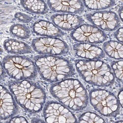
- Immunohistochemical staining of human colon shows distinct luminal membranous positivity in glandular cells.

- Submitted by
- Atlas Antibodies (provider)
- Main image

- Experimental details
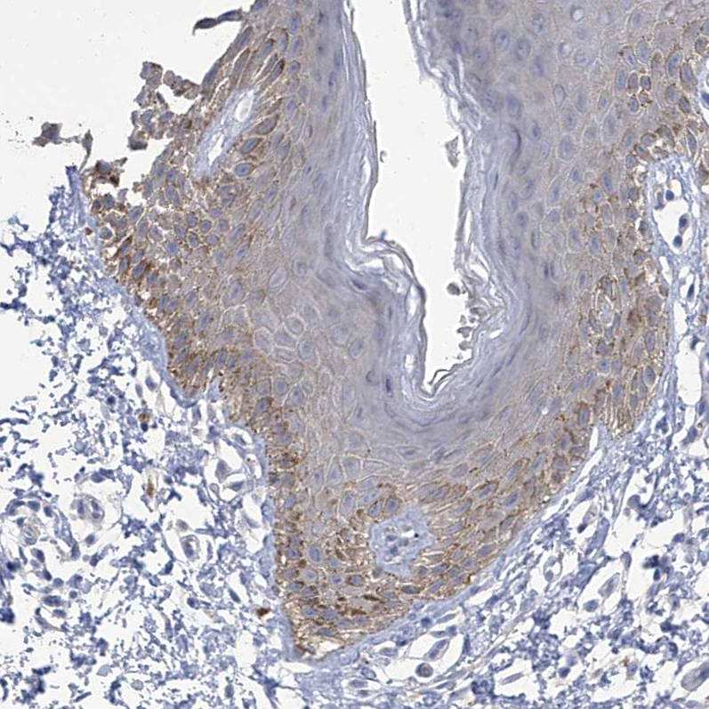
- Immunohistochemical staining of human skin shows weak to moderate cytoplasmic positivity in epidermal cells.
- Sample type
- HUMAN
- Submitted by
- Atlas Antibodies (provider)
- Main image
- Experimental details
- Immunohistochemical staining of human fallopian tube shows weak to moderate positivity in luminal membrane in glandular cells.
- Sample type
- HUMAN

- Submitted by
- Atlas Antibodies (provider)
- Main image

- Experimental details
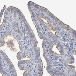
- Immunohistochemical staining of human colon shows weak to moderate positivity in luminal membrane in glandular cells.
- Sample type
- HUMAN

- Submitted by
- Atlas Antibodies (provider)
- Main image

- Experimental details
- Immunohistochemical staining of human cerebellum shows moderate positivity in neuropil.
- Sample type
- HUMAN

- Submitted by
- Atlas Antibodies (provider)
- Main image

- Experimental details
- Immunohistochemical staining of human kidney shows weak to moderate membranous positivity in cells in tubules.
- Sample type
- HUMAN
