 Explore
 Validate
 Learn
Learn Western blot
 Immunocytochemistry
Antibody data
- Antibody Data
- Antigen structure
- References [0]
- Comments [0]
- Validations
- Western blot [1]
- Immunohistochemistry [2]
- Flow cytometry [1]
Submit
Validation data
Reference
Comment
Report error
- Product number
- AP11348PU-N - Provider product page

- Provider
- Acris Antibodies GmbH
- Proper citation
- Acris Antibodies GmbH Cat#AP11348PU-N, RRID:AB_1749527
- Product name
- anti Cadherin-4 (N-term)
- Antibody type
- Polyclonal
- Antigen
- KLH conjugated synthetic peptide between 182~211 amino acids from the N-terminal region of human CDH4
- Reactivity
- Human
- Host
- Rabbit
- Vial size
- 0.4 ml
- Concentration
- lot specific
No comments: Submit comment
Supportive validation
- Submitted by
- Acris Antibodies GmbH (provider)
- Main image

- Experimental details
- Western blot analysis of anti-CDH4 Antibody (N-term) (RB13654) in HepG2 cell line lysates (35ug/lane). CDH4 (arrow) was detected using the purified Pab (1:60 dilution).

Supportive validation
- Submitted by
- Acris Antibodies GmbH (provider)
- Main image

- Experimental details
- Immunofluorescence analysis of CDH4 Antibody (N-term) cat.-No AP11348PU-N with paraffin-embedded human brain tissue . 0.025 mg/ml primary antibody was followed by FITC-conjugated goat anti-rabbit lgG (whole molecule). FITC emits green fluorescence.Red counterstaining is PI.

- Submitted by
- Acris Antibodies GmbH (provider)
- Main image

- Experimental details
- Formalin-fixed and paraffin-embedded human brain tissue reacted with CDH4 antibody (N-term) Cat.-No AP11348PU-N, which was peroxidase-conjugated to the secondary antibody, followed by DAB staining. This data demonstrates the use of this antibody for immunohistochemistry; clinical relevance has not been evaluated.

Supportive validation
- Submitted by
- Acris Antibodies GmbH (provider)
- Main image
- Experimental details
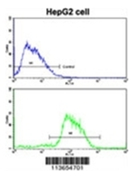
- Flow cytometric analysis of HepG2 cells using CDH4 Antibody (N-term) Cat.-No AP11348PU-N (bottom histogram) compared to a negative control cell (top histogram). FITC-conjugated goat-anti-rabbit secondary antibodies were used for the analysis.
