Explore
 Validate
 Learn
LearnPA5-32086
antibody from Invitrogen Antibodies
Targeting: TC2N
C14orf47, C2CD1, FLJ36557, MTAC2D1, Tac2-N
 Western blot
Antibody data
- Antibody Data
- Antigen structure
- References [1]
- Comments [0]
- Validations
- Western blot [1]
- Other assay [2]
Submit
Validation data
Reference
Comment
Report error
- Product number
- PA5-32086 - Provider product page

- Provider
- Invitrogen Antibodies
- Product name
- TC2N Polyclonal Antibody
- Antibody type
- Polyclonal
- Antigen
- Recombinant protein fragment
- Description
- Recommended positive controls: A549.
- Concentration
- 1 mg/mL
Submitted references Tac2-N serves an oncogenic role and promotes drug resistance in human gastric cancer cells.
Shen L, Zhang P, Wang J, Ji P
Experimental and therapeutic medicine 2020 Nov;20(5):113
Experimental and therapeutic medicine 2020 Nov;20(5):113
No comments: Submit comment
Supportive validation
- Submitted by
- Invitrogen Antibodies (provider)
- Main image

- Experimental details
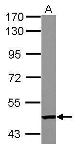
- Western Blot using TC2N Polyclonal Antibody (Product # PA5-32086). Sample (30 µg of whole cell lysate). Lane A: A549 . 7.5% SDS PAGE. TC2N Polyclonal Antibody (Product # PA5-32086) diluted at 1:1,000.
Supportive validation
- Submitted by
- Invitrogen Antibodies (provider)
- Main image

- Experimental details
- Figure 2 TC2N expression levels are upregulated in gastric cancer cell lines. The normal gastric mucosa cell line GES1 and gastric cancer cell line AGS were used to analyze the (A) mRNA and (B) protein expression levels of TC2N using reverse transcription-quantitative PCR and western blotting, respectively. (C) Semi-quantification of the expression levels presented in part (B). *** P

- Submitted by
- Invitrogen Antibodies (provider)
- Main image

- Experimental details
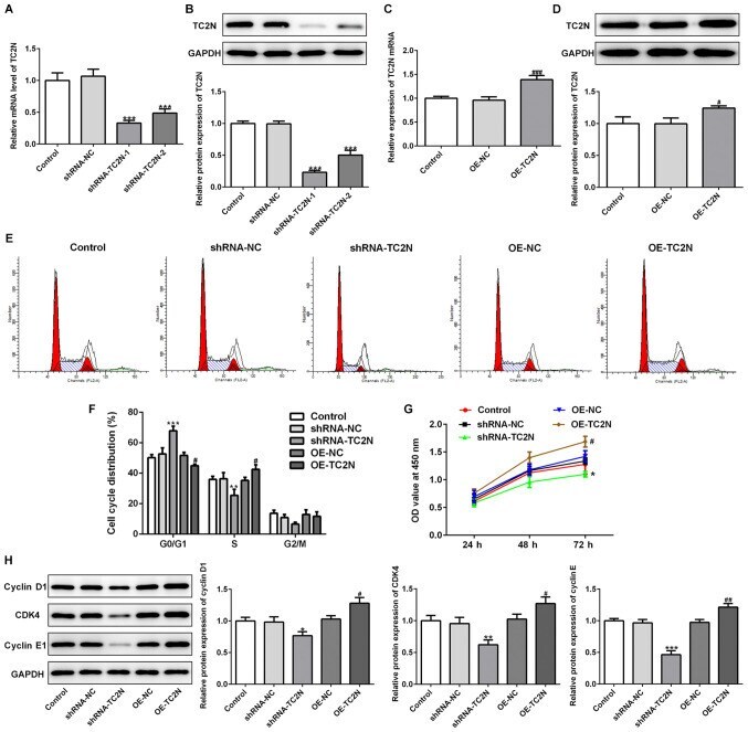
- Figure 3 TC2N regulates the cell proliferation of AGS cells. To determine the function of TC2N in gastric cancer, AGS cells were transfected with shRNA-TC2N and the (A) mRNA and (B) protein expression levels of TC2N were analyzed using RT-qPCR and western blotting, respectively. AGS cells were transfected with OE-TC2N and the (C) mRNA and (D) protein expression levels of TC2N were determined using RT-qPCR and western blotting, respectively. (E) Cell cycle distribution was analyzed using flow cytometric analysis. (F) Quantification of cell cycle distribution presented in part (E). (G) Cell Counting Kit-8 assay was performed to determine the cell proliferation rate. (H) Protein expression levels of cyclin D1, CDK4 and cyclin E1 were analyzed using western blotting. * P