Explore
 Validate
 Learn
Learn Western blot
Antibody data
- Antibody Data
- Antigen structure
- References [2]
- Comments [0]
- Validations
- Western blot [3]
- Immunocytochemistry [2]
- Immunohistochemistry [3]
Submit
Validation data
Reference
Comment
Report error
- Product number
- GTX108271 - Provider product page

- Provider
- GeneTex
- Proper citation
- GeneTex Cat#GTX108271, RRID:AB_1950232
- Product name
- eRF1 antibody
- Antibody type
- Polyclonal
- Reactivity
- Human, Mouse, Rat
- Host
- Rabbit
Submitted references Large-Scale Proteomic Identification of Targets of Cellular miR-197 Downregulated by Enterovirus A71.
Fip1 regulates mRNA alternative polyadenylation to promote stem cell self-renewal.
Tang WF, Huang RT, Chien KY, Tang P, Horng JT
Journal of proteome research 2019 Jan 4;18(1):449-460
Journal of proteome research 2019 Jan 4;18(1):449-460
Fip1 regulates mRNA alternative polyadenylation to promote stem cell self-renewal.
Lackford B, Yao C, Charles GM, Weng L, Zheng X, Choi EA, Xie X, Wan J, Xing Y, Freudenberg JM, Yang P, Jothi R, Hu G, Shi Y
The EMBO journal 2014 Apr 16;33(8):878-89
The EMBO journal 2014 Apr 16;33(8):878-89
No comments: Submit comment
Supportive validation
- Submitted by
- GeneTex (provider)
- Main image

- Experimental details
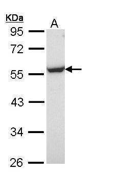
- Sample (30 ug of whole cell lysate) A: Molt-4 (GTX27912) 10% SDS PAGE GTX108271 diluted at 1:1000
- Validation comment
- WB
- Submitted by
- GeneTex (provider)
- Main image
- Experimental details
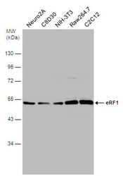
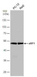
- Various whole cell extracts (30 £gg) were separated by 10% SDS-PAGE, and the membrane was blotted with eRF1 antibody (GTX108271) diluted at 1:500.
- Submitted by
- GeneTex (provider)
- Main image
- Experimental details
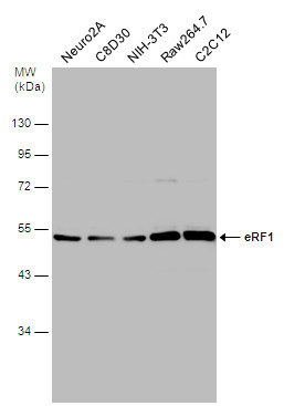
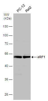
- Various whole cell extracts (30 £gg) were separated by 10% SDS-PAGE, and the membrane was blotted with eRF1 antibody (GTX108271) diluted at 1:500.
Supportive validation
- Submitted by
- GeneTex (provider)
- Main image
- Experimental details
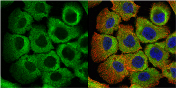
- eRF1 antibody detects eRF1 protein at cytoplasm by immunofluorescent analysis.Sample: HeLa cells were fixed in 4% paraformaldehyde at RT for 15 min.Green: eRF1 protein stained by eRF1 antibody (GTX108271) diluted at 1:500.Red: phalloidin, a cytoskeleton marker, stained by phalloidin (invitrogen, A12380) diluted at 1:200.Blue: Hoechst 33342 staining.

- Submitted by
- GeneTex (provider)
- Main image

- Experimental details
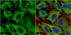
- eRF1 antibody detects eRF1 protein at cytoplasm by immunofluorescent analysis.Sample: A431 cells were fixed in 4% paraformaldehyde at RT for 15 min.Green: eRF1 protein stained by eRF1 antibody (GTX108271) diluted at 1:500.Red: alpha Tubulin, a cytoskeleton marker, stained by alpha Tubulin antibody [GT114] (GTX628802) diluted at 1:1000.Blue: Hoechst 33342 staining.
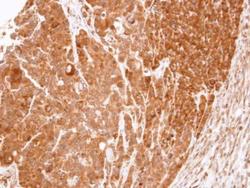
Supportive validation
- Submitted by
- GeneTex (provider)
- Main image
- Experimental details
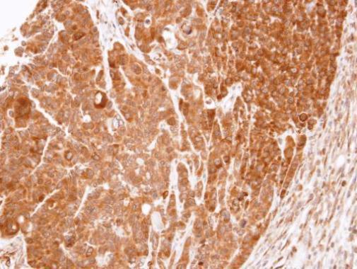
- Immunohistochemical analysis of paraffin-embedded SW480 xenograft, using eRF1(GTX108271) antibody at 1:100 dilution.
- Submitted by
- GeneTex (provider)
- Main image

- Experimental details
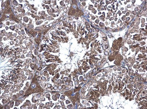
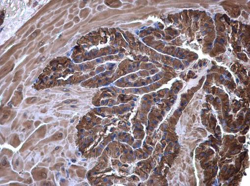
- eRF1 antibody detects eRF1 protein at cytoplasm on mouse prostate by immunohistochemical analysis. Sample: Paraffin-embedded mouse prostate. eRF1 antibody (GTX108271) diluted at 1:500.
- Submitted by
- GeneTex (provider)
- Main image
- Experimental details
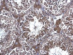
- eRF1 antibody detects eRF1 protein at cytoplasm on mouse testis by immunohistochemical analysis. Sample: Paraffin-embedded mouse testis. eRF1 antibody (GTX108271) diluted at 1:500.