 Explore
 Validate
 Learn
Learn Western blot
 Immunoprecipitation
Antibody data
- Antibody Data
- Antigen structure
- References [9]
- Comments [0]
- Validations
- Western blot [1]
- ELISA [1]
- Immunocytochemistry [1]
- Immunohistochemistry [1]
Submit
Validation data
Reference
Comment
Report error
- Product number
- H00009467-M01 - Provider product page

- Provider
- Novus Biologicals
- Proper citation
- Novus Cat#H00009467-M01, RRID:AB_2286068
- Product name
- Mouse Monoclonal SH3BP5 Antibody
- Antibody type
- Monoclonal
- Description
- Ascites. SH3BP5 - SH3-domain binding protein 5 (BTK-associated)
- Reactivity
- Human, Mouse, Rat
- Host
- Mouse
- Isotype
- IgG
- Vial size
- 0.1 mg
- Storage
- Aliquot and store at -20C or -80C. Avoid freeze-thaw cycles.
Submitted references N-n-Butyl Haloperidol Iodide Ameliorates Oxidative Stress in Mitochondria Induced by Hypoxia/Reoxygenation through the Mitochondrial c-Jun N-Terminal Kinase/Sab/Src/Reactive Oxygen Species Pathway in H9c2 Cells.
Sab concentrations indicate chemotherapeutic susceptibility in ovarian cancer cell lines.
Sab mediates mitochondrial dysfunction involved in imatinib mesylate-induced cardiotoxicity.
Detrusor induction of miR-132/212 following bladder outlet obstruction: association with MeCP2 repression and cell viability.
Sub-chronic administration of LY294002 sensitizes cervical cancer cells to chemotherapy by enhancing mitochondrial JNK signaling.
JNK interaction with Sab mediates ER stress induced inhibition of mitochondrial respiration and cell death.
Blocking c-Jun N-terminal kinase (JNK) translocation to the mitochondria prevents 6-hydroxydopamine-induced toxicity in vitro and in vivo.
Inhibition of JNK mitochondrial localization and signaling is protective against ischemia/reperfusion injury in rats.
Mitochondrial c-Jun N-terminal kinase (JNK) signaling initiates physiological changes resulting in amplification of reactive oxygen species generation.
Chu Q, Zhang Y, Zhong S, Gao F, Chen Y, Wang B, Zhang Z, Cai W, Li W, Zheng F, Shi G
Oxidative medicine and cellular longevity 2019;2019:7417561
Oxidative medicine and cellular longevity 2019;2019:7417561
Sab concentrations indicate chemotherapeutic susceptibility in ovarian cancer cell lines.
Paudel I, Hernandez SM, Portalatin GM, Chambers TP, Chambers JW
The Biochemical journal 2018 Nov 15;475(21):3471-3492
The Biochemical journal 2018 Nov 15;475(21):3471-3492
Sab mediates mitochondrial dysfunction involved in imatinib mesylate-induced cardiotoxicity.
Chambers TP, Santiesteban L, Gomez D, Chambers JW
Toxicology 2017 May 1;382:24-35
Toxicology 2017 May 1;382:24-35
Detrusor induction of miR-132/212 following bladder outlet obstruction: association with MeCP2 repression and cell viability.
Sadegh MK, Ekman M, Krawczyk K, Svensson D, Göransson O, Dahan D, Nilsson BO, Albinsson S, Uvelius B, Swärd K
PloS one 2015;10(1):e0116784
PloS one 2015;10(1):e0116784
Sub-chronic administration of LY294002 sensitizes cervical cancer cells to chemotherapy by enhancing mitochondrial JNK signaling.
Chambers TP, Portalatin GM, Paudel I, Robbins CJ, Chambers JW
Biochemical and biophysical research communications 2015 Aug 7;463(4):538-44
Biochemical and biophysical research communications 2015 Aug 7;463(4):538-44
JNK interaction with Sab mediates ER stress induced inhibition of mitochondrial respiration and cell death.
Win S, Than TA, Fernandez-Checa JC, Kaplowitz N
Cell death & disease 2014 Jan 9;5:e989
Cell death & disease 2014 Jan 9;5:e989
Blocking c-Jun N-terminal kinase (JNK) translocation to the mitochondria prevents 6-hydroxydopamine-induced toxicity in vitro and in vivo.
Chambers JW, Howard S, LoGrasso PV
The Journal of biological chemistry 2013 Jan 11;288(2):1079-87
The Journal of biological chemistry 2013 Jan 11;288(2):1079-87
Inhibition of JNK mitochondrial localization and signaling is protective against ischemia/reperfusion injury in rats.
Chambers JW, Pachori A, Howard S, Iqbal S, LoGrasso PV
The Journal of biological chemistry 2013 Feb 8;288(6):4000-11
The Journal of biological chemistry 2013 Feb 8;288(6):4000-11
Mitochondrial c-Jun N-terminal kinase (JNK) signaling initiates physiological changes resulting in amplification of reactive oxygen species generation.
Chambers JW, LoGrasso PV
The Journal of biological chemistry 2011 May 6;286(18):16052-62
The Journal of biological chemistry 2011 May 6;286(18):16052-62
No comments: Submit comment
Supportive validation
- Submitted by
- Novus Biologicals (provider)
- Main image
- Experimental details
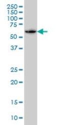
- Western Blot: SH3BP5 Antibody (2B3) [H00009467-M01] - SH3BP5 monoclonal antibody (M01), clone 2B3 Analysis of SH3BP5 expression in A-431.

Supportive validation
- Submitted by
- Novus Biologicals (provider)
- Main image
- Experimental details
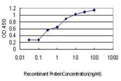
- ELISA: SH3BP5 Antibody (2B3) [H00009467-M01] - Detection limit for recombinant GST tagged SH3BP5 is approximately 0.03ng/ml as a capture antibody.

Supportive validation
- Submitted by
- Novus Biologicals (provider)
- Main image

- Experimental details
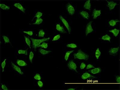
- Immunocytochemistry/Immunofluorescence: SH3BP5 Antibody (2B3) [H00009467-M01] - Analysis of monoclonal antibody to SH3BP5 on HeLa cell. Antibody concentration 10 ug/ml.
Supportive validation
- Submitted by
- Novus Biologicals (provider)
- Main image
- Experimental details
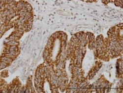
- Immunohistochemistry-Paraffin: SH3BP5 Antibody (2B3) [H00009467-M01] - Analysis of monoclonal antibody to SH3BP5 on formalin-fixed paraffin-embedded human colon adenocarcinoma. Antibody concentration 1 ug/ml.
