 Explore
 Validate
 Learn
Learn Western blot
Antibody data
- Antibody Data
- Antigen structure
- References [0]
- Comments [0]
- Validations
- Western blot [3]
- Immunocytochemistry [2]
- Flow cytometry [2]
Submit
Validation data
Reference
Comment
Report error
- Product number
- MA3-085 - Provider product page

- Provider
- Invitrogen Antibodies
- Product name
- FXN Monoclonal Antibody (NPM-1B2)
- Antibody type
- Monoclonal
- Antigen
- Other
- Description
- MA3-085 detects Frataxin from human and mouse samples. MA3-085 has been successfully used in Western blot, immunofluorescence, Flow Cytometry, and ELISA applications.
- Reactivity
- Human, Mouse
- Host
- Mouse
- Isotype
- IgG
- Antibody clone number
- NPM-1B2
- Vial size
- 50 μL
- Concentration
- Conc. Not Determined
- Storage
- -20°C, Avoid Freeze/Thaw Cycles
No comments: Submit comment
Supportive validation
- Submitted by
- Invitrogen Antibodies (provider)
- Main image
- Experimental details
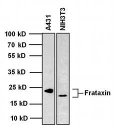
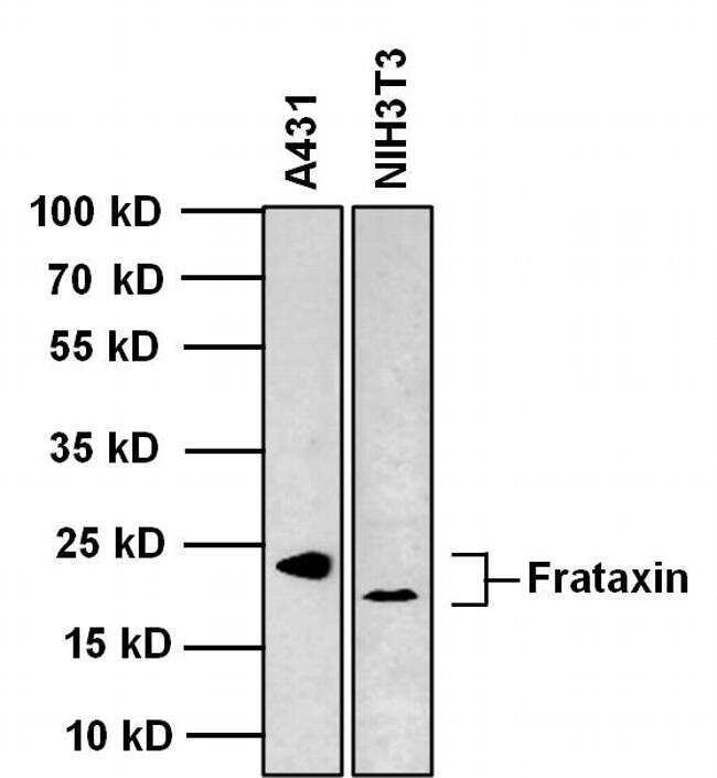
- Western blot analysis of Frataxin was performed by loading 20 µg of the indicated whole cell lysates and 5 µL of PageRuler Plus Prestained Protein Ladder (Product # 26619) per well onto a 4-20% Tris-Glycine polyacrylamide gel (Product # WT4202BX10). Proteins were transferred to a nitrocellulose membrane using the G2 Blotter (Product # 62288), and blocked with 5% Milk in TBST for 1 hour at room temperature. Frataxin was detected at 23 kDa (human) and 19 kDa (mouse) using a Frataxin mouse monoclonal antibody (Product # MA3-085) at a dilution of 1:500 in blocking buffer for 1 hour at room temperature on a rocking platform, followed by a Goat anti-Mouse IgG (H+L) Superclonal™ Secondary Antibody, HRP conjugate (Product # A28177) at a dilution of 1:1000 for at least 30 minutes at room temperature. Chemiluminescent detection was performed using SuperSignal West Pico substrate (Product # 34078) and the myECL Imager (Product # 62236).
- Submitted by
- Invitrogen Antibodies (provider)
- Main image
- Experimental details
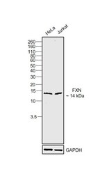
- Western Blot was performed using Anti-FXN Monoclonal Antibody (NPM-1B2) (Product # MA3-085) and a 14 kDa band corresponding to Frataxin, mitochondrial was observed across cell lines tested. Membrane enriched extracts (30 µg lysate) of HeLa (Lane 1) and Jurkat (Lane 2) were electrophoresed using Novex™ 16% Tricine Protein Gel (Product # EC6695BOX). Resolved proteins were then transferred onto a Nitrocellulose membrane (Product # IB23001) by iBlot® 2 Dry Blotting System (Product # IB21001). The Blot was probed with the primary antibody (1:1000 dilution) and detected by chemiluminescence with Goat anti-Mouse IgG (H+L) Superclonal™ Recombinant Secondary Antibody, HRP (Product # A28177, 1:4000 dilution) using the iBright FL 1000 (Product # A32752). Chemiluminescent detection was performed using SuperSignal™ West Dura Extended Duration Substrate (Product # 34076).

- Submitted by
- Invitrogen Antibodies (provider)
- Main image
- Experimental details
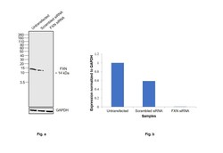
- Knockdown of Frataxin, mitochondrial was achieved by transfecting HeLa with Frataxin, mitochondrial specific siRNAs (Silencer® select Product # S530862, S5362). Western Blot analysis (Fig. a) was performed using Membrane enriched extracts from the Frataxin, mitochondrial knockdown cells (lane 3), non-targeting scrambled siRNA transfected cells (lane 2) and untransfected cells (lane 1). The Blot was probed with FXN Monoclonal Antibody (NPM-1B2) (Product # MA3-085, 1:1000 dilution ) and Goat anti-Mouse IgG (H+L) Superclonal™ Recombinant Secondary Antibody, HRP (Product # A28177, 1:4000 dilution). Densitometric analysis of this western Blot is shown in histogram (Fig. b). Decrease in signal upon siRNA mediated knock down confirms that antibody is specific to Frataxin, mitochondrial.

Supportive validation
- Submitted by
- Invitrogen Antibodies (provider)
- Main image
- Experimental details
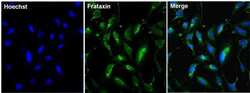
- Immunofluorescent analysis of Frataxin (green) in U2OS cells. The cells were fixed with 4% paraformaldehyde for 15 minutes, permeabilized with 0.1% Triton X-100 in PBS for 15 minutes, and blocked with 3% BSA in PBS (Product # 37525) for 30 minutes at room temperature. Cells were stained with a Frataxin mouse monoclonal antibody (Product # MA3-085) at a dilution of 1:500 in staining buffer for 1 hour at room temperature, and then incubated with a Goat anti-Mouse IgG Superclonal™ Secondary Antibody, Alexa Fluor 488 conjugate (Product # A28175) at a dilution of 1:1000 for 1 hour at room temperature. Nuclei (blue) were counterstained with Hoechst 33342 dye (Product # 62249). Images were taken on a Thermo Scientific ToxInsight Instrument at 20X magnification.
- Submitted by
- Invitrogen Antibodies (provider)
- Main image
- Experimental details
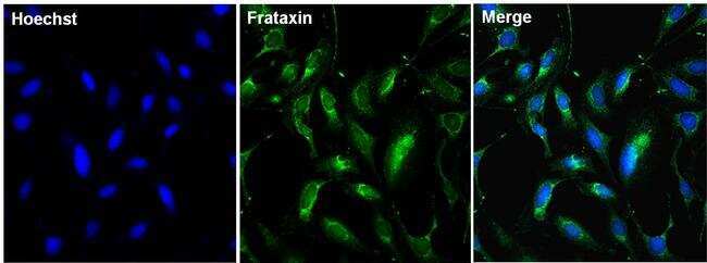
- Immunofluorescent analysis of Frataxin (green) in U2OS cells. The cells were fixed with 4% paraformaldehyde for 15 minutes, permeabilized with 0.1% Triton X-100 in PBS for 15 minutes, and blocked with 3% BSA in PBS (Product # 37525) for 30 minutes at room temperature. Cells were stained with a Frataxin mouse monoclonal antibody (Product # MA3-085) at a dilution of 1:500 in staining buffer for 1 hour at room temperature, and then incubated with a Goat anti-Mouse IgG Superclonal™ Secondary Antibody, Alexa Fluor 488 conjugate (Product # A28175) at a dilution of 1:1000 for 1 hour at room temperature. Nuclei (blue) were counterstained with Hoechst 33342 dye (Product # 62249). Images were taken on a Thermo Scientific ToxInsight Instrument at 20X magnification.

Supportive validation
- Submitted by
- Invitrogen Antibodies (provider)
- Main image
- Experimental details
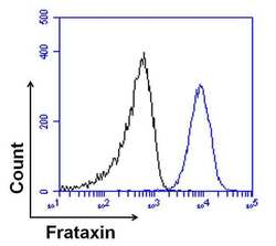
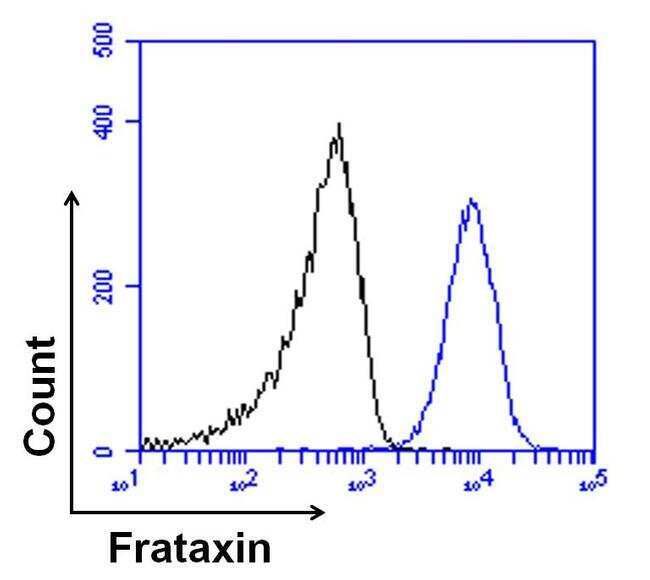
- Flow cytometry analysis of Frataxin was done on U2OS cells. Cells were fixed, permeabilized and stained with a Frataxin mouse monoclonal antibody (Product # MA3-081, blue histogram) at a dilution of 1:100. After incubation of the primary antibody on ice for an hour, the cells were stained with a Goat anti-mouse IgG Secondary Antibody, DyLight 680 conjugate (Product # 35519) at a dilution of 1:50 for 1 hour on ice. A representative 10,000 cells were acquired for each sample. The black histogram represents unstained control cells.
- Submitted by
- Invitrogen Antibodies (provider)
- Main image
- Experimental details
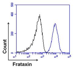
- Flow cytometry analysis of Frataxin was done on U2OS cells. Cells were fixed, permeabilized and stained with a Frataxin mouse monoclonal antibody (Product # MA3-081, blue histogram) at a dilution of 1:100. After incubation of the primary antibody on ice for an hour, the cells were stained with a Goat anti-mouse IgG Secondary Antibody, DyLight 680 conjugate (Product # 35519) at a dilution of 1:50 for 1 hour on ice. A representative 10,000 cells were acquired for each sample. The black histogram represents unstained control cells.
