Explore
 Validate
 Learn
LearnNBP2-24683
antibody from Novus Biologicals
Targeting: STING1
ERIS, FLJ38577, MITA, MPYS, NET23, STING, TMEM173
 Western blot
 Immunocytochemistry
Antibody data
- Antibody Data
- Antigen structure
- References [15]
- Comments [0]
- Validations
- Western blot [2]
- Immunohistochemistry [4]
- Flow cytometry [3]
Submit
Validation data
Reference
Comment
Report error
- Product number
- NBP2-24683 - Provider product page

- Provider
- Novus Biologicals
- Product name
- Rabbit Polyclonal STING/TMEM173 Antibody
- Antibody type
- Polyclonal
- Description
- Peptide affinity purified.
- Reactivity
- Human, Mouse, Canine, Simian
- Host
- Rabbit
- Isotype
- IgG
- Vial size
- 0.1 mg
- Concentration
- 1.0 mg/ml
- Storage
- Store at 4C short term. Aliquot and store at -20C long term. Avoid freeze-thaw cycles.
Submitted references Pharmacological STING Activation Is a Potential Alternative to Overcome Drug-Resistance in Melanoma.
STING couples with PI3K to regulate actin reorganization during BCR activation.
STING activation reprograms tumor vasculatures and synergizes with VEGFR2 blockade.
RAD51 interconnects between DNA replication, DNA repair and immunity.
Sensing of HSV-1 by the cGAS-STING pathway in microglia orchestrates antiviral defence in the CNS.
Antitumor Activity of cGAMP via Stimulation of cGAS-cGAMP-STING-IRF3 Mediated Innate Immune Response.
Evasion of innate cytosolic DNA sensing by a gammaherpesvirus facilitates establishment of latent infection.
Identification of a negative feedback loop between cyclic di-GMP-induced levels of IFI16 and p202 cytosolic DNA sensors and STING.
Inflammatory cytokines break down intrinsic immunological tolerance of human primary keratinocytes to cytosolic DNA.
Listeria monocytogenes induces IFNβ expression through an IFI16-, cGAS- and STING-dependent pathway.
Inhibition of dengue and chikungunya virus infections by RIG-I-mediated type I interferon-independent stimulation of the innate antiviral response.
IFI16 senses DNA forms of the lentiviral replication cycle and controls HIV-1 replication.
Proteasomal degradation of herpes simplex virus capsids in macrophages releases DNA to the cytosol for recognition by DNA sensors.
Nuclear IFI16 induction of IRF-3 signaling during herpesviral infection and degradation of IFI16 by the viral ICP0 protein.
A role for DNA-dependent activator of interferon regulatory factor in the recognition of herpes simplex virus type 1 by glial cells.
Chipurupalli S, Ganesan R, Dhanabal SP, Kumar MS, Robinson N
Frontiers in oncology 2020;10:758
Frontiers in oncology 2020;10:758
STING couples with PI3K to regulate actin reorganization during BCR activation.
Jing Y, Dai X, Yang L, Kang D, Jiang P, Li N, Cheng J, Li J, Miller H, Ren B, Gong Q, Yin W, Liu Z, Mattila PK, Ning Q, Sun J, Yu B, Liu C
Science advances 2020 Apr;6(17):eaax9455
Science advances 2020 Apr;6(17):eaax9455
STING activation reprograms tumor vasculatures and synergizes with VEGFR2 blockade.
Yang H, Lee WS, Kong SJ, Kim CG, Kim JH, Chang SK, Kim S, Kim G, Chon HJ, Kim C
The Journal of clinical investigation 2019 Jul 25;129(10):4350-4364
The Journal of clinical investigation 2019 Jul 25;129(10):4350-4364
RAD51 interconnects between DNA replication, DNA repair and immunity.
Bhattacharya S, Srinivasan K, Abdisalaam S, Su F, Raj P, Dozmorov I, Mishra R, Wakeland EK, Ghose S, Mukherjee S, Asaithamby A
Nucleic acids research 2017 May 5;45(8):4590-4605
Nucleic acids research 2017 May 5;45(8):4590-4605
Sensing of HSV-1 by the cGAS-STING pathway in microglia orchestrates antiviral defence in the CNS.
Reinert LS, Lopušná K, Winther H, Sun C, Thomsen MK, Nandakumar R, Mogensen TH, Meyer M, Vægter C, Nyengaard JR, Fitzgerald KA, Paludan SR
Nature communications 2016 Nov 10;7:13348
Nature communications 2016 Nov 10;7:13348
Antitumor Activity of cGAMP via Stimulation of cGAS-cGAMP-STING-IRF3 Mediated Innate Immune Response.
Li T, Cheng H, Yuan H, Xu Q, Shu C, Zhang Y, Xu P, Tan J, Rui Y, Li P, Tan X
Scientific reports 2016 Jan 12;6:19049
Scientific reports 2016 Jan 12;6:19049
Evasion of innate cytosolic DNA sensing by a gammaherpesvirus facilitates establishment of latent infection.
Sun C, Schattgen SA, Pisitkun P, Jorgensen JP, Hilterbrand AT, Wang LJ, West JA, Hansen K, Horan KA, Jakobsen MR, O'Hare P, Adler H, Sun R, Ploegh HL, Damania B, Upton JW, Fitzgerald KA, Paludan SR
Journal of immunology (Baltimore, Md. : 1950) 2015 Feb 15;194(4):1819-31
Journal of immunology (Baltimore, Md. : 1950) 2015 Feb 15;194(4):1819-31
Identification of a negative feedback loop between cyclic di-GMP-induced levels of IFI16 and p202 cytosolic DNA sensors and STING.
Panchanathan R, Liu H, Xin D, Choubey D
Innate immunity 2014 Oct;20(7):751-9
Innate immunity 2014 Oct;20(7):751-9
Inflammatory cytokines break down intrinsic immunological tolerance of human primary keratinocytes to cytosolic DNA.
Chiliveru S, Rahbek SH, Jensen SK, Jørgensen SE, Nissen SK, Christiansen SH, Mogensen TH, Jakobsen MR, Iversen L, Johansen C, Paludan SR
Journal of immunology (Baltimore, Md. : 1950) 2014 Mar 1;192(5):2395-404
Journal of immunology (Baltimore, Md. : 1950) 2014 Mar 1;192(5):2395-404
Listeria monocytogenes induces IFNβ expression through an IFI16-, cGAS- and STING-dependent pathway.
Hansen K, Prabakaran T, Laustsen A, Jørgensen SE, Rahbæk SH, Jensen SB, Nielsen R, Leber JH, Decker T, Horan KA, Jakobsen MR, Paludan SR
The EMBO journal 2014 Aug 1;33(15):1654-66
The EMBO journal 2014 Aug 1;33(15):1654-66
Inhibition of dengue and chikungunya virus infections by RIG-I-mediated type I interferon-independent stimulation of the innate antiviral response.
Olagnier D, Scholte FE, Chiang C, Albulescu IC, Nichols C, He Z, Lin R, Snijder EJ, van Hemert MJ, Hiscott J
Journal of virology 2014 Apr;88(8):4180-94
Journal of virology 2014 Apr;88(8):4180-94
IFI16 senses DNA forms of the lentiviral replication cycle and controls HIV-1 replication.
Jakobsen MR, Bak RO, Andersen A, Berg RK, Jensen SB, Tengchuan J, Laustsen A, Hansen K, Ostergaard L, Fitzgerald KA, Xiao TS, Mikkelsen JG, Mogensen TH, Paludan SR
Proceedings of the National Academy of Sciences of the United States of America 2013 Nov 26;110(48):E4571-80
Proceedings of the National Academy of Sciences of the United States of America 2013 Nov 26;110(48):E4571-80
Proteasomal degradation of herpes simplex virus capsids in macrophages releases DNA to the cytosol for recognition by DNA sensors.
Horan KA, Hansen K, Jakobsen MR, Holm CK, Søby S, Unterholzner L, Thompson M, West JA, Iversen MB, Rasmussen SB, Ellermann-Eriksen S, Kurt-Jones E, Landolfo S, Damania B, Melchjorsen J, Bowie AG, Fitzgerald KA, Paludan SR
Journal of immunology (Baltimore, Md. : 1950) 2013 Mar 1;190(5):2311-9
Journal of immunology (Baltimore, Md. : 1950) 2013 Mar 1;190(5):2311-9
Nuclear IFI16 induction of IRF-3 signaling during herpesviral infection and degradation of IFI16 by the viral ICP0 protein.
Orzalli MH, DeLuca NA, Knipe DM
Proceedings of the National Academy of Sciences of the United States of America 2012 Oct 30;109(44):E3008-17
Proceedings of the National Academy of Sciences of the United States of America 2012 Oct 30;109(44):E3008-17
A role for DNA-dependent activator of interferon regulatory factor in the recognition of herpes simplex virus type 1 by glial cells.
Furr SR, Chauhan VS, Moerdyk-Schauwecker MJ, Marriott I
Journal of neuroinflammation 2011 Aug 12;8:99
Journal of neuroinflammation 2011 Aug 12;8:99
No comments: Submit comment
Supportive validation
- Submitted by
- Novus Biologicals (provider)
- Main image
- Experimental details
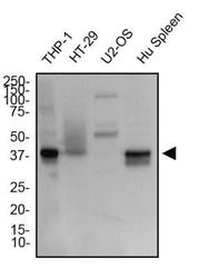
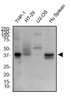
- Western Blot: STING/TMEM173 Antibody [NBP2-24683] - Total protein from THP-1, HT-29, U2OS cells and human spleen was separated on a 12% gel by SDS-PAGE, transferred to PVDF membrane and blocked in 5% non-fat milk in TBST. The membrane was probed with 2.0 ug/ml STING/TMEM173 Antibody in 1% non-fat milk in TBST and detected with an anti-rabbit HRP secondary antibody using chemiluminescence.
- Submitted by
- Novus Biologicals (provider)
- Main image

- Experimental details
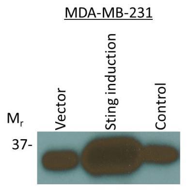
- Western Blot: STING/TMEM173 Antibody [NBP2-24683] - STING/TMEM173 expression was induced in human breast MDA-MB-231 cells followed by Western blotting using STING/TMEM173 Antibody antibody (1:1000). Only one specific band at an apparent molecular mass of 37 kDa was observed. Image from verified customer review.
Supportive validation
- Submitted by
- Novus Biologicals (provider)
- Main image

- Experimental details
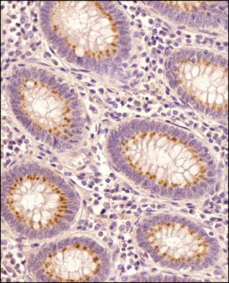
- Immunohistochemistry-Paraffin: STING/TMEM173 Antibody [NBP2-24683] - Human colon cancer tissue section using STING/TMEM173 Antibody at 1:100 dilution with detection employing HRP-conjugated secondary antibody. The signal was developed using DAB reagent and the nuclei were counterstained with hematoxylin. The antibody generated very weak cytoplasmic staining in columnar epithelial cells with a very strong signal in the secretory/goblet cells.
- Submitted by
- Novus Biologicals (provider)
- Main image
- Experimental details
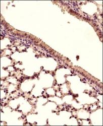
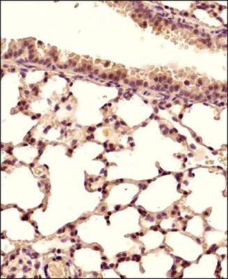
- Immunohistochemistry-Paraffin: STING/TMEM173 Antibody [NBP2-24683] - Mouse lung tissue section using STING/TMEM173 Antibody at 1:150 dilution with detection employing HRP-conjugated secondary antibody. The signal was developed using DAB reagent and the nuclei were counterstained with hematoxylin. The antibody generated mainly a cytoplasmic staining in the bronchiolar and alveolar epithelial cells.
- Submitted by
- Novus Biologicals (provider)
- Main image
- Experimental details
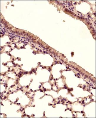
- Immunohistochemistry-Paraffin: STING/TMEM173 Antibody [NBP2-24683] - Mouse lung tissue section using STING antibody at 1:150 dilution with detection employing HRP-conjugated secondary antibody. The signal was developed using DAB reagent and the nuclei were counterstained with hematoxylin. The antibody generated mainly a cytoplasmic staining in the bronchiolar and alveolar epithelial cells.
- Submitted by
- Novus Biologicals (provider)
- Main image
- Experimental details
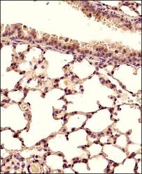
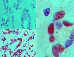
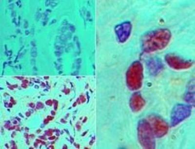
- Immunohistochemistry-Paraffin: STING/TMEM173 Antibody [NBP2-24683] - Analysis of STING in FFPE human breast tumor using an isotype control (top left) and this antibody (bottom left, right) at 1:100 dilution.
Supportive validation
- Submitted by
- Novus Biologicals (provider)
- Main image
- Experimental details
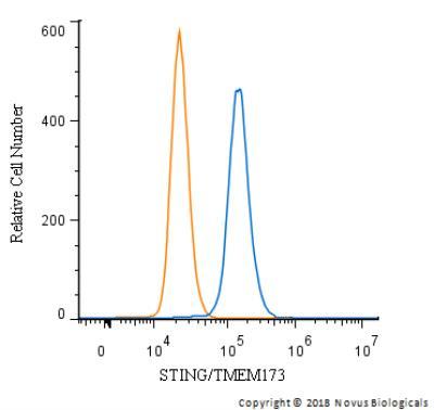
- Flow Cytometry: STING/TMEM173 Antibody [NBP2-24683] - An intracellular stain was performed on THP-1 cells with STING/TMEM173 Antibody and a matched isotype control. Cells were fixed with 4% PFA and then permeablized with 0.1% saponin. Cells were incubated in an antibody dilution of 2.5 ug/mL for 30 minutes at room temperature, followed by Rabbit IgG (H+L) Cross-Adsorbed Secondary Antibody.
- Submitted by
- Novus Biologicals (provider)
- Main image
- Experimental details
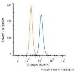
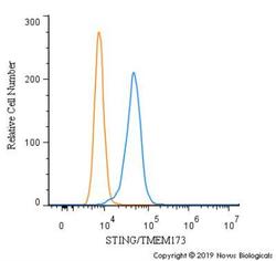
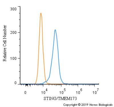
- Flow Cytometry: STING/TMEM173 Antibody [NBP2-24683] - An intracellular stain was performed on RH30 cells with STING/TMEM173 Antibody (blue) and a matched isotype control (orange). Cells were fixed with 4% PFA and then permeabilized with 0.1% saponin. Cells were incubated in an antibody dilution of 2.5 ug/mL for 30 minutes at room temperature, followed by Rabbit IgG (H+L) Cross-Adsorbed Secondary Antibody.
- Submitted by
- Novus Biologicals (provider)
- Main image
- Experimental details
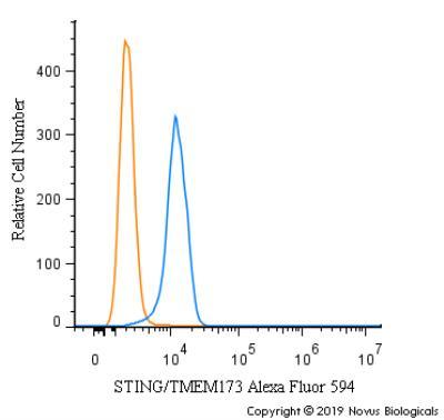
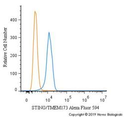
- Flow Cytometry: STING/TMEM173 Antibody [NBP2-24683] - An intracellular stain was performed on U937 cells with NBP2-24683AF594 (blue) and a matched isotype control (orange). Cells were fixed with 4% PFA and then permeabilized with 0.1% saponin. Cells were incubated in an antibody dilution of 2.5 ug/mL for 30 minutes at room temperature. Both antibodies were conjugated to Alexa Fluor 594.