Explore
 Validate
 Learn
Learn Western blot
Antibody data
- Antibody Data
- Antigen structure
- References [1]
- Comments [0]
- Validations
- Western blot [1]
- Immunocytochemistry [1]
- Other assay [1]
Submit
Validation data
Reference
Comment
Report error
- Product number
- 702076 - Provider product page

- Provider
- Invitrogen Antibodies
- Product name
- SLC6A4 Recombinant Rabbit Monoclonal Antibody (16H2L7)
- Antibody type
- Monoclonal
- Antigen
- Synthetic peptide
- Reactivity
- Human, Mouse, Rat
- Host
- Rabbit
- Isotype
- IgG
- Antibody clone number
- 16H2L7
- Vial size
- 100 µg
- Concentration
- 0.5 mg/mL
- Storage
- Store at 4°C short term. For long term storage, store at -20°C, avoiding freeze/thaw cycles.
Submitted references The effect of citalopram treatment on amyloid-β precursor protein processing and oxidative stress in human hNSC-derived neurons.
Elsworthy RJ, Crowe JA, King MC, Dunleavy C, Fisher E, Ludlam A, Parri HR, Hill EJ, Aldred S
Translational psychiatry 2022 Jul 18;12(1):285
Translational psychiatry 2022 Jul 18;12(1):285
No comments: Submit comment
Supportive validation
- Submitted by
- Invitrogen Antibodies (provider)
- Main image

- Experimental details
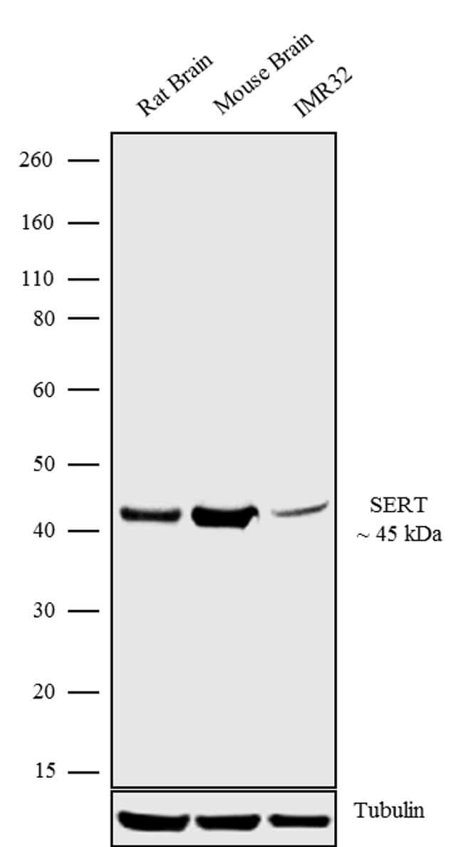
- Western blot analysis was performed on whole cell extracts (30 µg lysate) of Rat Brain (Lane 1), Mouse Brain (Lane 2) and IMR32 (Lane 3). The blots were probed with Anti-SERT Recombinant Rabbit Monoclonal Antibody (Product # 702076, 1-2 µg/mL) and detected by chemiluminescence using Goat anti-Rabbit IgG (H+L) Superclonal™ Secondary Antibody, HRP conjugate (Product # A27036, 0.4 µg/mL, 1:2500 dilution). A 45 kDa band corresponding to SERT was observed across the cell lines tested. Known quantity of protein samples were electrophoresed using Novex® NuPAGE® 4-12% Bis-Tris gel (Product # NP0321BOX), XCell SureLock™ Electrophoresis System (Product # EI0002) and Novex® Sharp Pre-Stained Protein Standard (Product # LC5800). Resolved proteins were then transferred onto a nitrocellulose membrane with iBlot® Dry Blotting System (Product # IB21001). The membrane was probed with the relevant primary and secondary Antibody following blocking with 5% skimmed milk. Chemiluminescent detection was performed using Pierce™ ECL Western blotting Substrate (Product # 32106).
Supportive validation
- Submitted by
- Invitrogen Antibodies (provider)
- Main image

- Experimental details
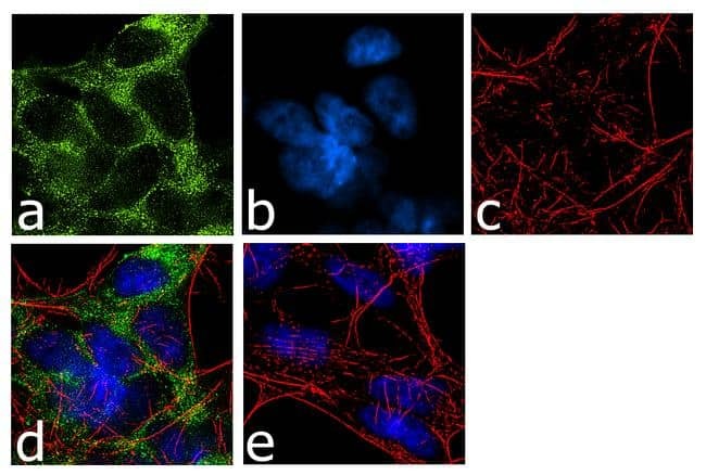
- For immunofluorescence analysis, IMR-32 cells were fixed and permeabilized for detection of endogenous SERT using Anti- SERT Recombinant Rabbit Monoclonal Antibody (Product # 702076, 2 µg/mL) and labeled with Goat anti-Rabbit IgG (H+L) Superclonal™ Secondary Antibody, Alexa Fluor® 488 conjugate (Product # A27034, 1:2000). Panel a) shows representative cells that were stained for detection and localization of SERT protein (green), Panel b) is stained for nuclei (blue) using SlowFade® Gold Antifade Mountant with DAPI (Product # S36938). Panel c) represents cytoskeletal F-actin staining using Alexa Fluor® 555 Rhodamine Phalloidin (Product # R415, 1:300). Panel d) is a composite image of Panels a, b and c clearly demonstrating membrane localization of SERT. Panel e) represents control cells with no primary antibody to assess background. The images were captured at 60X magnification.
Supportive validation
- Submitted by
- Invitrogen Antibodies (provider)
- Main image
- Experimental details
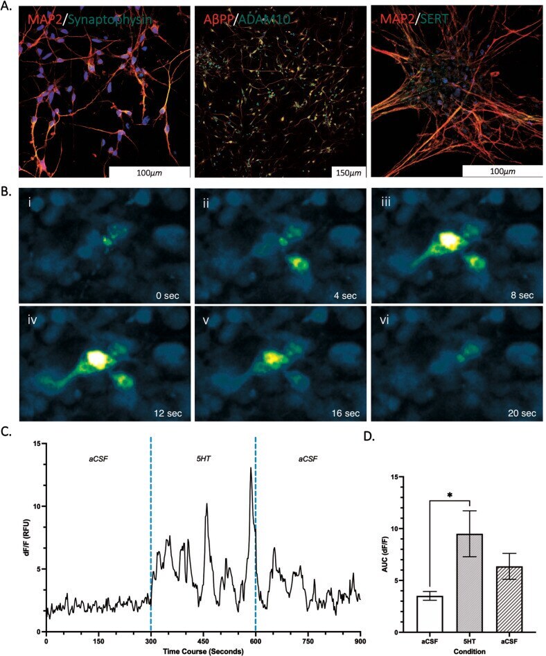
- Representative microscopy images of cultured cells with ICC staining (A) and calcium imaging (B & C) ( n = 3). A Mature neuronal staining positive for MAP2, Synaptophysin, SERT, ADAM10 and AbetaPP (AX0018, d45) with nucleus staining for DAPI (blue). B PSEN1 (L286V) fluoro-4-am calcium transient measured in neurons (day 42). C Signal intensity was used to calculate neuronal activity (DF/F) under basal (aCSF), during stimulation with 5HT and a wash off period (aCSF) in 5-min intervals ( n = 3). D Area under the curve plotted for each condition to analyse neuronal activity (3ROIs, n = 3). Data normality (p > 0.05) and S.D. Variation (p < 0.05) confirmed assumptions for ANOVA analysis with multiple comparison corrections applied. *Significantly different to treatment condition ( p < 0.05).